")
Tak1 Antikörper (C-9): sc-7967
- Tak1 Antikörper C-9 ist ein Maus monoklonales IgG2a κ Tak1 Antikörper, verwendet in 66 wissenschaftlichen Veröffentlichungen, in einer Menge von 200 µg/ml
- gegen Aminosäuren 1-579 aufgezogen, die die vollständige Länge Tak1 (TGFβ-aktivierte Kinase) von mouse darstellen
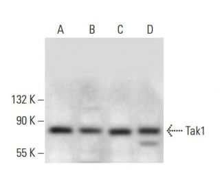
- Tak1 Antikörper (C-9) ist empfohlen für die Detektion von Tak1 aus der Spezies mouse, rat und human per WB, IP, IF und ELISA
- Anti-Tak1 Antikörper (C-9) ist erhältlich als Konjugat mit Agarose für IP; HRP für WB, IHC(P) und ELISA; und entweder mit Phycoerythrin oder FITC für IF, IHC(P) und FCM
- auch erhältlich als Konjugat mit Alexa Fluor® 488, Alexa Fluor® 546, Alexa Fluor® 594 oder Alexa Fluor® 647 für IF, IHC(P) und FCM
- auch erhältlich als Konjugat mit Alexa Fluor® 680 oder Alexa Fluor® 790 für WB (NIR), IF und FCM
- m-IgG Fc BP-HRP und m-IgGκ BP-HRP sind die bevorzugten sekundären Nachweisreagenzien für Tak1 Antikörper (C-9) für WB-Anwendungen. Diese Reagenzien werden jetzt in Bündeln mit Tak1 Antikörper (C-9) angeboten(siehe Bestellinformationen unten).

Direktverknüpfungen
Der Tak1-Antikörper (C-9) ist ein monoklonaler IgG2a-Kappa-Kappa-Leichtketten-Antikörper der Maus, der gegen die Aminosäuren 1-579 von Maus-Tak1 (TGFβ-aktivierte Kinase 1), auch bekannt als MAP3K7 (Mitogen-aktivierte Protein-Kinase-Kinase-Kinase 7), gerichtet ist. Der monoklonale Antikörper (C-9) gegen Tak1 weist das Tak1-Protein von Mäusen, Ratten und Menschen durch Western Blot (WB), Immunpräzipitation (IP), Immunfluoreszenz (IF) und Enzyme-linked Immunosorbent Assay (ELISA) nach. Tak1 ist eine Serin/Threonin-Kinase, die eine entscheidende Rolle bei der Vermittlung zellulärer Reaktionen auf Zytokine wie TGFβ und Interleukin-1 (IL-1) spielt, indem sie nachgeschaltete NF-κB- und MAP-Kinase-Signalwege aktiviert, die für Entzündungen, Immunantworten, das Überleben von Zellen und die Apoptose unerlässlich sind. Eine Fehlregulation der Tak1-Funktion ist mit verschiedenen pathologischen Zuständen verbunden, darunter Krebs und entzündliche Erkrankungen, was Tak1 zu einem wichtigen Ziel für die biomedizinische Forschung macht. Der monoklonale Antikörper Tak1 (C-9) ermöglicht die Untersuchung wichtiger Signalwege und ihrer Rolle bei Krankheitsmechanismen. Der monoklonale Antikörper Tak1 (C-9) ist in nicht konjugierter Form und in mehreren konjugierten Formen erhältlich, darunter Agarose, HRP, PE, FITC und mehrere Alexa Fluor®-Konjugate, was vielseitige Anwendungen in der Forschung ermöglicht.
Alexa Fluor® ist ein Markenzeichen von Molecular Probes Inc., OR., USA
LI-COR® und Odyssey® sind Markenzeichen von LI-COR Biosciences
Tak1 Antikörper (C-9) Literaturhinweise:
- Proteinkinase-Kaskaden bei der Kontrolle des meiotischen und mitotischen Zellzyklus. | Pelech, SL., et al. 1990. Biochem Cell Biol. 68: 1297-330. PMID: 2085430
- Signaltransduktion von der Membran zum Zytoplasma: Wachstumsfaktoren und membrangebundene Onkogenprodukte erhöhen die Raf-1-Phosphorylierung und die damit verbundene Proteinkinaseaktivität. | Morrison, DK., et al. 1988. Proc Natl Acad Sci U S A. 85: 8855-9. PMID: 3057494
- Insulin-stimulierte Mikrotubuli-assoziierte Proteinkinase wird in vivo an Tyrosin und Threonin phosphoryliert. | Ray, LB. and Sturgill, TW. 1988. Proc Natl Acad Sci U S A. 85: 3753-7. PMID: 3287375
- TAK1 vermittelt neuronale Pyroptose bei frühen Hirnverletzungen nach Subarachnoidalblutungen. | Xu, P., et al. 2021. J Neuroinflammation. 18: 188. PMID: 34461942
- Charakterisierung des murinen A-raf, eines neuen Onkogens, das mit dem v-raf Onkogen verwandt ist. | Huleihel, M., et al. 1986. Mol Cell Biol. 6: 2655-62. PMID: 3491291
- Die Rolle von TAK1 bei der RANKL-induzierten Osteoklastogenese. | Jianwei, W., et al. 2022. Calcif Tissue Int. 111: 1-12. PMID: 35286417
- TAK1-Mangel fördert Leberschäden und Tumorentstehung durch Ferroptose und Makrophagen-cGAS-STING-Signalübertragung. | Su, W., et al. 2023. JHEP Rep. 5: 100695. PMID: 36968217
- TAK1 ist eine essentielle Kinase für den STING-Transport. | Ma, M., et al. 2023. Mol Cell. 83: 3885-3903.e5. PMID: 37832545
- KSR: ein neuer Akteur im RAS-Signalweg. | Downward, J. 1995. Cell. 83: 831-4. PMID: 8521506
- KSR, eine neue Proteinkinase, die für die RAS-Signaltransduktion erforderlich ist. | Therrien, M., et al. 1995. Cell. 83: 879-88. PMID: 8521512
- Das ksr-1-Gen von C. elegans kodiert eine neue Raf-verwandte Kinase, die an der Ras-vermittelten Signaltransduktion beteiligt ist. | Sundaram, M. and Han, M. 1995. Cell. 83: 889-901. PMID: 8521513
- Identifizierung eines Mitglieds der MAPKKK-Familie als potenzieller Vermittler der TGF-beta-Signaltransduktion. | Yamaguchi, K., et al. 1995. Science. 270: 2008-11. PMID: 8533096
Bestellinformation
| Produkt | Katalog # | EINHEIT | Preis | ANZAHL | Favoriten | |
Tak1 Antikörper (C-9) | sc-7967 | 200 µg/ml | $322.00 | |||
Tak1 (C-9): m-IgG Fc BP-HRP Bundle | sc-528190 | 200 µg Ab; 10 µg BP | $361.00 | |||
Tak1 (C-9): m-IgGκ BP-HRP Bundle | sc-520512 | 200 µg Ab, 40 µg BP | $361.00 | |||
Tak1 Antikörper (C-9) AC | sc-7967 AC | 500 µg/ml, 25% agarose | $424.00 | |||
Tak1 Antikörper (C-9) HRP | sc-7967 HRP | 200 µg/ml | $322.00 | |||
Tak1 Antikörper (C-9) FITC | sc-7967 FITC | 200 µg/ml | $336.00 | |||
Tak1 Antikörper (C-9) PE | sc-7967 PE | 200 µg/ml | $349.00 | |||
Tak1 Antikörper (C-9) Alexa Fluor® 488 | sc-7967 AF488 | 200 µg/ml | $364.00 | |||
Tak1 Antikörper (C-9) Alexa Fluor® 546 | sc-7967 AF546 | 200 µg/ml | $364.00 | |||
Tak1 Antikörper (C-9) Alexa Fluor® 594 | sc-7967 AF594 | 200 µg/ml | $364.00 | |||
Tak1 Antikörper (C-9) Alexa Fluor® 647 | sc-7967 AF647 | 200 µg/ml | $364.00 | |||
Tak1 Antikörper (C-9) Alexa Fluor® 680 | sc-7967 AF680 | 200 µg/ml | $364.00 | |||
Tak1 Antikörper (C-9) Alexa Fluor® 790 | sc-7967 AF790 | 200 µg/ml | $364.00 |