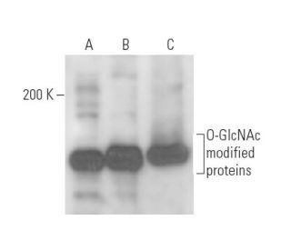
Date published: 2025-9-11

00800 4573 8000

SCBT Portrait Logo
Seach Input

O-GlcNAc Antikörper (CTD110.6): sc-59623

4.7(10)
Produkt bewertenBitte stellen Sie eine Frage

Datenblätter
  • O-GlcNAc Antikörper CTD110.6 ist ein Maus monoklonales IgM κ O-GlcNAc Antikörper, verwendet in 71 wissenschaftlichen Veröffentlichungen, in einer Menge von 200 µg/ml
  • gegen O-GlcNAc mit Serin-O-verknüpftem N-Acetylglucosamin
  • Empfohlen für die Detektion von Ser-O-GlcNAc and Thr-O-GlcNAc aus der Spezies a broad range of species, including mammals, insects, worms, plants und filamentous fungi per WB und IP; keine Kreuzreaktivität mit peptide determinants or other closely-related carbohydrate antigens
  • Als Direktkonjugat zur Detektion von O-GlcNAc wird O-GlcNAc (RL2): sc-59624 angeboten; Primärantikörper konjugiert mit AC, HRP, FITC, PE, Alexa Fluor® 488, 594, 647, 680 und 790.
  • Bitte kontaktieren Sie uns um ein 10 µg GRATIS Sample vom O-GlcNAc (CTD110.6): sc-59623 zu erhalten.
  • Aktuell testen wir noch unsere Sekundärantikörper um das beste Bindeprotein für diesen Primärantikörper O-GlcNAc (CTD110.6) zu finden. Kontaktieren Sie uns bitte, wenn Sie Fragen hierzu haben sollten.

Direktverknüpfungen

Siehe auch...

Der O-GlcNAc-Antikörper (CTD110.6) ist ein monoklonaler Maus IgM κ O-GlcNAc-Antikörper (auch als O-verknüpfter β-N-Acetylglucosamin-Antikörper, O-β-GlcNAc-Antikörper, O-verknüpfter GlcNAc-Antikörper, Serin-Hydroxyl-O-GlcNAc-Antikörper, Threonin-Hydroxyl-O-GlcNAc-Antikörper oder O-GlcNAcylation-spezifischer Antikörper bezeichnet), der das O-GlcNAc-Protein einer breiten Palette von Arten, einschließlich Säugetieren, Insekten, Würmern, Pflanzen und fadenförmigen Pilzen, mittels WB und IP detektiert. Der O-GlcNAc-Antikörper (CTD110.6) ist als nicht konjugierter Anti-O-GlcNAc-Antikörper erhältlich. O-GlcNAc (O-verknüpftes N-Acetylglucosamin) ist eine Form der Protein-Glykosylierung, die ausschließlich im Kern und Cytoplasma eukaryotischer Zellen vorkommt. Viele Proteine werden an ihren Serin- und Threonin-Hydroxylgruppen durch die Anbindung von O-GlcNAc modifiziert. Proteine, die den Transport in und aus dem Kernporen regulieren, sind stark O-GlcNAcyliert. Phosphorylierte O-GlcNAc-Proteine bilden reversibel multimere Komplexe mit anderen Proteinen und diese Assoziationen werden häufig durch Phosphorylierung reguliert. O-GlcNAc-Proteine können eine Schlüsselrolle bei der Pathogenese von Tumoren und verschiedenen Krebszellen spielen. O-GlcNAc-Reste regulieren die Assemblierung des Preinitiation-Komplexes und sind daher wichtig für die Transkription. Cytoskelett- und Membran-O-GlcNAc-Proteine erhalten die Zellform der Erythrozyten und regulieren die Degradation von Proteinen, die für Läsionen in der Alzheimer-Krankheit verantwortlich sind.

Ausschließlich für Forschungszwecke. Nicht für diagnostische oder therapeutische Zwecke bestimmt.

Alexa Fluor® ist ein Markenzeichen von Molecular Probes Inc., OR., USA

LI-COR® und Odyssey® sind Markenzeichen von LI-COR Biosciences

O-GlcNAc Antikörper (CTD110.6) Literaturhinweise:

  1. Das O-GlcNAc-Transferase-Gen befindet sich auf dem X-Chromosom und ist für die Lebensfähigkeit embryonaler Stammzellen und die Ontogenese der Maus von wesentlicher Bedeutung.  |  Shafi, R., et al. 2000. Proc Natl Acad Sci U S A. 97: 5735-9. PMID: 10801981
  2. Lokalisierung der O-GlcNAc-Transferase und O-GlcNAc-modifizierter Proteine in der Kleinhirnrinde der Ratte.  |  Akimoto, Y., et al. 2003. Brain Res. 966: 194-205. PMID: 12618343
  3. Glykosylierung von Kern- und Zytoplasmaproteinen. Reinigung und Charakterisierung einer Uridindiphospho-N-Acetylglucosamin:Polypeptid Beta-N-Acetylglucosaminyltransferase.  |  Haltiwanger, RS., et al. 1992. J Biol Chem. 267: 9005-13. PMID: 1533623
  4. Identifizierung eines geheimen Wirkstoffs als O-GlcNAc-Transferase, die an der Infektion mit dem Pflaumenpockenvirus beteiligt ist.  |  Chen, D., et al. 2005. J Virol. 79: 9381-7. PMID: 16014901
  5. Ein Hochdurchsatz-Assay für O-GlcNAc-Transferase erkennt primäre Sequenzpräferenzen in Peptidsubstraten.  |  Leavy, TM. and Bertozzi, CR. 2007. Bioorg Med Chem Lett. 17: 3851-4. PMID: 17531489
  6. Phosphoinositid-Signalisierung verbindet O-GlcNAc-Transferase mit Insulinresistenz.  |  Yang, X., et al. 2008. Nature. 451: 964-9. PMID: 18288188
  7. O-GlcNAc-Transferase: Strukturelle Eigenschaften, katalytischer Mechanismus und niedermolekulare Inhibitoren.  |  Ju Kim, E. 2020. Chembiochem. 21: 3026-3035. PMID: 32406185
  8. Fortschritte bei der chemischen Untersuchung der Protein-O-GlcNAc-Glykosylierung: strukturelle Rolle und molekulare Mechanismen.  |  Saha, A., et al. 2021. Chem Soc Rev. 50: 10451-10485. PMID: 34338261
  9. Integration von O-GlcNAc in Stressreaktionswege.  |  Fahie, KMM., et al. 2022. Cells. 11: PMID: 36359905
  10. Die O-GlcNAc-Modifikation von GSDMD schwächt die LPS-induzierte Pyroptose von Endothelzellen ab.  |  Yu, F., et al. 2024. Inflamm Res. 73: 5-17. PMID: 37962578
  11. Dynamische Glykosylierung von Kern- und Zytosolproteinen. Klonierung und Charakterisierung einer einzigartigen O-GlcNAc-Transferase mit mehreren Tetratricopeptid-Wiederholungen.  |  Kreppel, LK., et al. 1997. J Biol Chem. 272: 9308-15. PMID: 9083067
  12. Die O-gebundene GlcNAc-Transferase ist ein konserviertes nukleozytoplasmatisches Protein, das Tetratripeptidwiederholungen enthält.  |  Lubas, WA., et al. 1997. J Biol Chem. 272: 9316-24. PMID: 9083068

Bestellinformation

ProduktKatalog #EINHEITPreisANZAHLFavoriten

O-GlcNAc Antikörper (CTD110.6)

sc-59623
200 µg/ml
$316.00

I need specifically detect O-GlcNAcylation un plant proteim extractos. We found lack of specificity using CTD110.6. Employment of a secondary anti-IgM is mandatory? It could be conveniente to use RL2?

Gefragt von: Salamandra
Thank you for your question. It would be helpful if you could call us, allowing for a more interactive discussion of this and other related questions.
Beantwortet von: Technical Support
Veröffentlichungsdatum: 2021-07-23

I am planning to IP this modification after protein digestion to peptides and wonder if the antibody recognise digested peptides carrying this modifications and if you would expect some specificity for certain peptides?

Gefragt von: Paolo
Thank you for your question. Unfortunately I do not have any information regarding if this antibody can detect digested peptides.
Beantwortet von: Tech Service
Veröffentlichungsdatum: 2019-04-09

What is the best way to avoid background when working on mouse samples with this mouse monoclonal antibody?

Gefragt von: AbPolly
Thank you for your inquiry. Our new line of Mouse IgG binding proteins can help reduce non-specific staining with mouse samples. A complete list of available binding proteins is available on our website here: https://www.scbt.com/scbt/browse/support-products-mouse-igg-binding-proteins/_/N-ecrety You can also use our alternative O-GlcNAc antibody sc-59624 in a variety of direct conjugations to reduce non-specific staining with mouse samples.
Beantwortet von: Technical Service
Veröffentlichungsdatum: 2016-12-22
  • y_2025, m_8, d_28, h_5CST
  • bvseo_bulk, prod_bvqa, vn_bulk_3.0.42
  • cp_1, bvpage1
  • co_hasquestionsanswers, tq_3
  • loc_de_DE, sid_59623, prod, sort_[SortEntry(order=LAST_APPROVED_ANSWER_SUBMISSION_TIME, direction=DESCENDING)]
  • clientName_scbt
  • bvseo_sdk, java_sdk, bvseo-4.0.0
  • CLOUD, getContent, 119ms
  • QUESTIONS, PRODUCT
Rated 5 von 5 von aus goodvery nice,it work well in WB。Good specificity,nonspecific bands
Veröffentlichungsdatum: 2021-07-31
Rated 4 von 5 von aus clear signal in immunofluorescence, dirty in WBThis antibody worked well in immunofluorescence in cell culture and also in paraffin embedded human brain tissue. In western blot, it needs a lot of washes after primary antibody especially, usually the first time there's a dark smear everywhere and if exposure is less than 5 min there's no signal. Usually after re-application of the secondary the signal gets clearer, perhaps due to longer washing of primary. Primary seems to stick, but this is a problem of this clone, not of this product in particular. Here are the captions again for the vertical (tissue) and horizontal (cells) IF pictures and the western blot since they did not fit in the space given: Cells: Immunofluorescence for O-GlcNAc in human astrocytes. A) no primary antibody. B) DMSO treatment (control). C) 200 uM Voc OGT inhibitor treatment. D) 100 uM 9D OGA inhibitor treatment. Green: O-GlcNAc (Santa Cruz CTD110.6), Blue: DAPI. Primary antibody was used at 1:100 dilution overnight in 5% goat serum in PBS. Secondary antibody was Goat anti-mouse IgM Alexafluor 488 conjugate. Tissue: Immunofluorescence in human tissue. A) IF of formalin fixed paraffin embedded human brain tissue. Control patient in his 60s. B) Alzheimer’s disease patient in his 60s. Green: O-GlcNAc. Blue: DAPI. Tissues were reparaffinized, rehydrated, antigen retrieval was performed in boiling citrate buffer, free formaldehydes were blocked with sodium borohydride. Blocking and antibody incubations were done in 5% goat serum in PBS. Lipofuscin was blocked with Sudan black for 20 min. after DAPI incubation. Western blot: Western blot with human astrocyte lysates. Multiple samples were run on SDS-PAGE gels and transferred to PVDF membrane. Blocking was with 5% milk in TBST. Primary antibody was used at 1:100 dilution in 5% BSA in TBST. Secondary antibody (rabbit anti-mouse IgM HRP) was diluted 1:1000 in 5% milk in TBST. Membrane was washed 2x 5 mins then 30 min in TBST after primary and then 3x 5 min after secondary. Imaging was for 10 mins.
Veröffentlichungsdatum: 2020-12-30
Rated 5 von 5 von aus good result do WBWe used this antibody recenlty and following the struction choose the dilution 1:400(we feel it can diluted more),the band is right size,very good result.
Veröffentlichungsdatum: 2018-05-26
Rated 4 von 5 von aus Worked wellI bought antibody for pilot experiments and it woked well in Western. I will be happy to buy more for future work.
Veröffentlichungsdatum: 2018-02-16
Rated 5 von 5 von aus Really good !I used this antibody several times. It works well on human lung cells. even using a low dilution
Veröffentlichungsdatum: 2017-07-05
Rated 5 von 5 von aus Great for Western blotMonoclonal O-GlcNAc (CTD110.6) antibody has been validated by Western blot. Highly recommended!
Veröffentlichungsdatum: 2016-12-19
Rated 5 von 5 von aus Publishable WBPublishable WB.CoIP data with rat aortic smooth muscle cells - SCBT Publication Review
Veröffentlichungsdatum: 2015-04-10
Rated 4 von 5 von aus Dirty blot but produced bands at expectedDirty blot but produced bands at expected MW in HeLa nuclear extract and A549 whole cell lysate and mouse brain tissue extract. -SCBT QC
Veröffentlichungsdatum: 2015-03-26
  • y_2025, m_8, d_28, h_5
  • bvseo_bulk, prod_bvrr, vn_bulk_3.0.42
  • cp_1, bvpage1
  • co_hasreviews, tv_0, tr_10
  • loc_de_DE, sid_59623, prod, sort_[SortEntry(order=SUBMISSION_TIME, direction=DESCENDING)]
  • clientName_scbt
  • bvseo_sdk, java_sdk, bvseo-4.0.0
  • CLOUD, getReviews, 14ms
  • REVIEWS, PRODUCT
O-GlcNAc Antikörper (CTD110.6) wurde bewertet mit 4.7 von 5 von 10.
  • y_2025, m_8, d_28, h_5
  • bvseo_bulk, prod_bvrr, vn_bulk_3.0.42
  • cp_1, bvpage1
  • co_hasreviews, tv_0, tr_10
  • loc_de_DE, sid_59623, prod, sort_[SortEntry(order=SUBMISSION_TIME, direction=DESCENDING)]
  • clientName_scbt
  • bvseo_sdk, java_sdk, bvseo-4.0.0
  • CLOUD, getAggregateRating, 117ms
  • REVIEWS, PRODUCT