")
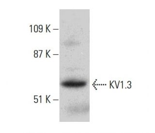
: sc-398855. Análisis Western blot de la expresión de KV1.3 en lisado de células Jurkat.")
: sc-398855. Análisis por Western blot de la expresión de KV1.3 en lisados de células 293T no transfectadas: sc-117752 (A), 293T transfectadas con KV1.3 humano: sc-159570 (B) y Jurkat (C).")
KV1.3 Anticuerpo (G-9): sc-398855
- KV1.3 Anticuerpo G-9 es un monoclonal de ratón IgG3 KV1.3 Anticuerpo, ver las 2 publicaciones, proporcionado como 200 µg/ml
- específico para un epítopo localizado entre los amino ácidos 535-556 dentro de C-terminal dominio citoplasmático de KV1.3 de origen human
- recomendado para detectarKV1.3 de mouse, rat y human origen, mediante WB, IP, IF y ELISA
- m-IgG Fc BP-HRP es el reactivo de detección secundario preferido para KV1.3 Anticuerpo (G-9) para aplicaciones WB. Este reactivo se ofrece ahora en un paquete con KV1.3 Anticuerpo (G-9)(ver información de pedido más abajo).

ENLACES RÁPIDOS
VER TAMBIÉN ....
El anticuerpo KV1.3 (G-9) es un anticuerpo monoclonal de ratón IgG3 que detecta la proteína KV1.3 de origen humano, de ratón y de rata mediante las técnicas de WB, IP, IF y ELISA. Este anticuerpo está disponible como anticuerpo anti-KV1.3 no conjugado. Los canales de potasio (K+) regulan la repolarización y la frecuencia de los potenciales de acción en neuronas, músculos y otras células excitables. La familia de genes KV codifica más de 30 genes que forman las subunidades de los canales de K+, y varían en sus propiedades de activación y permeación, distribución subcelular y patrones de expresión. Los canales KV funcionales se ensamblan como tetrameros que consisten en subunidades α formadoras de poros (KVα), que incluyen las proteínas KV1, KV2, KV3 y KV4, y subunidades accesorias o KVβ que modifican las propiedades de activación de las subunidades KVα coexpresadas. Existen diferencias en los patrones de tráfico, procesamiento biosintético y expresión en la superficie de las principales subunidades KV1 (KV1.1, KV1.2, KV1.4, KV1.5 y KV1.6) expresadas en el cerebro de rata y humano, lo que sugiere que las subunidades proteicas individuales están altamente reguladas para controlar el ensamblaje y formación de canales neuronales funcionales.
Alexa Fluor® es una marca registrada de Molecular Probes Inc., OR., USA
REIVEW LI-COR® y Odyssey® son marcas registradas de LI-COR Biosciences.
KV1.3 Anticuerpo (G-9) Referencias:
- Composición de subunidades de los canales Kv1 en el SNC humano. | Coleman, SK., et al. 1999. J Neurochem. 73: 849-58. PMID: 10428084
- La composición de subunidades determina la expresión superficial del canal de potasio Kv1. | Manganas, LN. and Trimmer, JS. 2000. J Biol Chem. 275: 29685-93. PMID: 10896669
- Los canales Kv1.3 facilitan la conexión entre el metabolismo y el flujo sanguíneo en el corazón. | Ohanyan, V., et al. 2017. Microcirculation. 24: PMID: 28504408
- Kv1.3 modula la neuroinflamación y la neurodegeneración en la enfermedad de Parkinson. | Sarkar, S., et al. 2020. J Clin Invest. 130: 4195-4212. PMID: 32597830
- Fisiología del canal de K+ Kv1.3 evaluada mediante modulación genética y farmacológica. | Varanita, T., et al. 2023. Physiology (Bethesda). 38: 0. PMID: 35998249
- El bloqueo del canal de potasio Kv1.3 inhibe la neuroinflamación mediada por la microglía en la epilepsia. | Zhang, X., et al. 2022. Int J Mol Sci. 23: PMID: 36499018
- Kv1.3 en el punto de mira para el tratamiento de enfermedades inmunitarias. | Navarro-Pérez, M., et al. 2024. Expert Opin Ther Targets. 28: 67-82. PMID: 38316438
- Modulación dependiente de KCNE4 de la farmacología de Kv1.3. | Sastre, D., et al. 2024. Biochem Pharmacol. 226: 116368. PMID: 38880360
- El canal de potasio cerebral Kv1.1: estudios in vitro e in vivo sobre el ensamblaje de subunidades y el procesamiento postraduccional. | Deal, KK., et al. 1994. J Neurosci. 14: 1666-76. PMID: 8126562
- Localización inmunohistoquímica de cinco miembros de las subunidades del canal Kv1: localizaciones subcelulares contrastadas y co-localizaciones neuronales específicas en cerebro de rata. | Veh, RW., et al. 1995. Eur J Neurosci. 7: 2189-205. PMID: 8563969
- Las subunidades beta promueven la expresión de la superficie de los canales K+ a través de efectos tempranos en la biosíntesis. | Shi, G., et al. 1996. Neuron. 16: 843-52. PMID: 8608002
- Asociación y colocalización de las subunidades beta Kvbeta1 y Kvbeta2 con las subunidades alfa Kv1 en complejos de canales K+ de cerebro de mamífero. | Rhodes, KJ., et al. 1997. J Neurosci. 17: 8246-58. PMID: 9334400
Información sobre pedidos
| Nombre del producto | Número de catálogo | UNIDAD | Precio | CANTIDAD | Favoritos | |
KV1.3 Anticuerpo (G-9) | sc-398855 | 200 µg/ml | $322.00 | |||
Paquete de KV1.3 (G-9): m-IgG Fc BP-HRP | sc-526332 | 200 µg Ab; 10 µg BP | $361.00 | |||
KV1.3 (G-9) péptido neutralizante | sc-398855 P | 100 µg/0.5 ml | $69.00 |