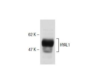
Date published: 2025-9-7

00800 4573 8000

SCBT Portrait Logo
Seach Input

HYAL1 Antikörper (1D10): sc-101340

4.6(7)
Produkt bewertenBitte stellen Sie eine Frage

Datenblätter
  • HYAL1 Antikörper 1D10 ist ein Maus monoklonales IgG1 κ HYAL1 Antikörper, verwendet in 8 wissenschaftlichen Veröffentlichungen, in einer Menge von 200 µg/ml
  • gezogen gegen die Aminosäuresequenz 1-366 von HYAL1 aus der Spezies human
  • HYAL1 Antikörper (1D10) ist empfohlen für die Detektion von HYAL1 aus der Spezies mouse, rat, human und hamster per WB, IP, IF und IHC(P); keine Kreuzreaktivität mit HYAL2 or HYAL3
  • Anti-HYAL1 Antikörper (1D10) ist erhältlich als Konjugat mit Agarose für IP; HRP für WB, IHC(P) und ELISA; und entweder mit Phycoerythrin oder FITC für IF, IHC(P) und FCM
  • auch erhältlich als Konjugat mit Alexa Fluor® 488, Alexa Fluor® 546, Alexa Fluor® 594 oder Alexa Fluor® 647 für IF, IHC(P) und FCM
  • auch erhältlich als Konjugat mit Alexa Fluor® 680 oder Alexa Fluor® 790 für WB (NIR), IF und FCM
  • Bitte kontaktieren Sie uns um ein 10 µg GRATIS Sample vom HYAL1 (1D10): sc-101340 zu erhalten.
  • m-IgG Fc BP-HRP und m-IgG1 BP-HRP sind die empfohlenen Detektionsreagenzien für HYAL1 Antikörper (1D10) für WB and IHC(P) Experimente. Diese Reagenzien werden nun auch als Bundle angeboten, zusammen mit HYAL1 Antikörper (1D10) (Bestellinformation unten stehend).

Direktverknüpfungen

Siehe auch...

Der HYAL1-Antikörper (1D10) ist ein monoklonaler Maus IgG1 κ HYAL1-Antikörper, der das HYAL1-Protein von Maus-, Ratte-, Mensch- und Hamster-Ursprung durch WB, IP, IF und IHC (P) detektiert. Der HYAL1-Antikörper (1D10) ist sowohl in nicht konjugierter Form als auch in mehreren konjugierten Formen des HYAL1-Antikörpers erhältlich, einschließlich Agarose, HRP, PE, FITC und mehreren Alexa Fluor®-Konjugaten. Hyaluronidasen (HAasen oder HYALs) sind eine Familie lysosomaler Enzyme, die für die Ausbreitung bakterieller Infektionen und von Toxinen in einer Vielzahl von Giften entscheidend sind. HYALs können auch an der Progression von Krebs beteiligt sein. Bei Menschen wurden sechs HYAL-Proteine identifiziert. Die meisten HYAL-Proteine spalten Hyaluronsäure (HA) ab, die in Körperflüssigkeiten, Geweben und der extrazellulären Matrix von Wirbeltiergeweben vorhanden ist. HA hält Gewebe hydriert, erhält den osmotischen Gleichgewicht und fördert die Zellproliferation, Differenzierung und Metastasierung. HA ist auch eine wichtige strukturelle Komponente von Knorpel und anderen Geweben und wirkt als Schmiermittel in Gelenken. HYAL1 ist eine 435 Aminosäure Hyaluronidase, die in mehreren Geweben exprimiert wird, insbesondere im Serum, und nicht im Gehirn exprimiert wird. HYAL1 spaltet HA in Fragmente, die die Angiogenese stimulieren. Die Expression von HYAL1 in verschiedenen Krebszellen kann eine Rolle bei der Regulation des Tumorwachstums und der Progression spielen.

Ausschließlich für Forschungszwecke. Nicht für diagnostische oder therapeutische Zwecke bestimmt.

Alexa Fluor® ist ein Markenzeichen von Molecular Probes Inc., OR., USA

LI-COR® und Odyssey® sind Markenzeichen von LI-COR Biosciences

HYAL1 Antikörper (1D10) Literaturhinweise:

  1. Die Charakterisierung der Genregion der murinen Hyaluronidase zeigt eine komplexe Organisation und Co-Transkription von Hyal1 mit den nachgeschalteten Genen Fus2 und Hyal3.  |  Shuttleworth, TL., et al. 2002. J Biol Chem. 277: 23008-18. PMID: 11929860
  2. Expression der Tumormarker Hyaluronsäure und Hyaluronidase (HYAL1) in Kopf-Hals-Tumoren.  |  Franzmann, EJ., et al. 2003. Int J Cancer. 106: 438-45. PMID: 12845686
  3. Vergleich des prognostischen Potenzials von Hyaluronsäure, Hyaluronidase (HYAL-1), CD44v6 und Mikrogefäßdichte bei Prostatakrebs.  |  Ekici, S., et al. 2004. Int J Cancer. 112: 121-9. PMID: 15305383
  4. Das Hyaluronidase-Gen HYAL1 ist beim Menschen auf Chromosom 3p21.2-p21.3 und bei der Maus auf Chromosom 9F1-F2 kartiert, einem konservierten Tumorsuppressor-Locus.  |  Csóka, AB., et al. 1998. Genomics. 48: 63-70. PMID: 9503017
  5. Unterschiedliche Selektivität von Hyaluronidase-Inhibitoren gegenüber sauren und basischen Hyaluronidasen.  |  Isoyama, T., et al. 2006. Glycobiology. 16: 11-21. PMID: 16166602

Bestellinformation

ProduktKatalog #EINHEITPreisANZAHLFavoriten

HYAL1 Antikörper (1D10)

sc-101340
200 µg/ml
$316.00

HYAL1 (1D10): m-IgG Fc BP-HRP Bundle

sc-527064
200 µg Ab; 10 µg BP
$354.00

HYAL1 (1D10): m-IgG1 BP-HRP Bundle

sc-532437
200 µg Ab; 20 µg BP
$354.00

HYAL1 Antikörper (1D10) AC

sc-101340 AC
500 µg/ml, 25% agarose
$416.00

HYAL1 Antikörper (1D10) HRP

sc-101340 HRP
200 µg/ml
$316.00

HYAL1 Antikörper (1D10) FITC

sc-101340 FITC
200 µg/ml
$330.00

HYAL1 Antikörper (1D10) PE

sc-101340 PE
200 µg/ml
$343.00

HYAL1 Antikörper (1D10) Alexa Fluor® 488

sc-101340 AF488
200 µg/ml
$357.00

HYAL1 Antikörper (1D10) Alexa Fluor® 546

sc-101340 AF546
200 µg/ml
$357.00

HYAL1 Antikörper (1D10) Alexa Fluor® 594

sc-101340 AF594
200 µg/ml
$357.00

HYAL1 Antikörper (1D10) Alexa Fluor® 647

sc-101340 AF647
200 µg/ml
$357.00

HYAL1 Antikörper (1D10) Alexa Fluor® 680

sc-101340 AF680
200 µg/ml
$357.00

HYAL1 Antikörper (1D10) Alexa Fluor® 790

sc-101340 AF790
200 µg/ml
$357.00

Hi, are the fluorophore-conjugated antibodies usable for intracellular staining and flow cytometry?

Gefragt von: jonn
Anti-HYAL1 Antibody (1D10) is available conjugated to Alexa Fluor® 488, Alexa Fluor® 546, Alexa Fluor® 594 or Alexa Fluor® 647 for WB (RGB), IF, IHC(P) and FCM, and for use with RGB fluorescent imaging systems, such as iBright™ FL1000, FluorChem™, Typhoon, Azure and other comparable systems
Beantwortet von: Technical Service
Veröffentlichungsdatum: 2024-05-07

Hi, I am concerning on the reactivity of this HYAL1 antibody with bovine hyaluronidases, especcially in bovine serum. Do you have information about that?

Gefragt von: kenny09
Thank you for your question. This antibody has not yet been tested in bovine samples and so that is not a recommended species at this time. We are still gathering data to show on our website. Check back for updates!
Beantwortet von: Tech Service
Veröffentlichungsdatum: 2019-12-06

What is the recommended secondary for this antibody?

Gefragt von: AbPolly
Thank you for your question. We recommend using one our exclusive Mouse IgG Binding Proteins as a secondary detection reagent. A complete list of available binding proteins is available on our website here: https://www.scbt.com/scbt/browse/support-products-mouse-igg-binding-proteins/_/N-ecrety
Beantwortet von: Technical Service
Veröffentlichungsdatum: 2017-02-02
  • y_2025, m_9, d_6, h_5CST
  • bvseo_bulk, prod_bvqa, vn_bulk_3.0.42
  • cp_1, bvpage1
  • co_hasquestionsanswers, tq_3
  • loc_de_DE, sid_101340, prod, sort_[SortEntry(order=LAST_APPROVED_ANSWER_SUBMISSION_TIME, direction=DESCENDING)]
  • clientName_scbt
  • bvseo_sdk, java_sdk, bvseo-4.0.0
  • CLOUD, getContent, 109ms
  • QUESTIONS, PRODUCT
Rated 4 von 5 von aus Ottimo Funzionamento!Abbiamo acquistato questo anticorpo un mese fa e l'abbiamo testa su rene umano. Performa molto bene!
Veröffentlichungsdatum: 2020-11-10
Rated 3 von 5 von aus Funktioniert in IHC für MausnierenWie im Bild funktioniert dieser AK in IHC für Mausnieren, allerdings haben wir Probleme, geeignete Zweit-AK zu finden, sodass wir bei weitem nicht so gute Ergebnisse wie im Musterbild haben. Wenn jemand Tipps hat, bitte posten.
Veröffentlichungsdatum: 2018-02-02
Rated 5 von 5 von aus Absolut überzeugendes ErgebnisIch habe den Antikörper von Santa Cruz verwendet, weil ich sowohl mit einem Polyklonalen als auch mit einem Monoklonalen Hyal-1 Antikörper nicht zufrieden war. Bzw. keine überzeugenden Ergebnisse erzielen konnte. Anders mit diesem Antikörper. Der Westernblot ergab eine spezifische Bande, so wie man es sich wünscht. Ich bin sehr zufrieden.
Veröffentlichungsdatum: 2017-02-28
Rated 5 von 5 von aus Worked in western blotWe used this antibody for a western blot and got a band at the correct molecular weight
Veröffentlichungsdatum: 2017-01-03
Rated 5 von 5 von aus Publishable IHC data with human lungPublishable IHC data with human lung - SCBT Publication Review
Veröffentlichungsdatum: 2015-02-22
Rated 5 von 5 von aus Produced great immunoperoxidase cytoplasmicProduced great immunoperoxidase cytoplasmic staining in formalin fixed, paraffin-embedded human kidney tissue. -SCBT QC
Veröffentlichungsdatum: 2015-02-18
Rated 5 von 5 von aus WB publishable data with human ASMCWB publishable data with human ASMC - SCBT Publication Review
Veröffentlichungsdatum: 2014-12-17
  • y_2025, m_9, d_6, h_5
  • bvseo_bulk, prod_bvrr, vn_bulk_3.0.42
  • cp_1, bvpage1
  • co_hasreviews, tv_0, tr_7
  • loc_de_DE, sid_101340, prod, sort_[SortEntry(order=SUBMISSION_TIME, direction=DESCENDING)]
  • clientName_scbt
  • bvseo_sdk, java_sdk, bvseo-4.0.0
  • CLOUD, getReviews, 10ms
  • REVIEWS, PRODUCT
HYAL1 Antikörper (1D10) wurde bewertet mit 4.6 von 5 von 7.
  • y_2025, m_9, d_6, h_5
  • bvseo_bulk, prod_bvrr, vn_bulk_3.0.42
  • cp_1, bvpage1
  • co_hasreviews, tv_0, tr_7
  • loc_de_DE, sid_101340, prod, sort_[SortEntry(order=SUBMISSION_TIME, direction=DESCENDING)]
  • clientName_scbt
  • bvseo_sdk, java_sdk, bvseo-4.0.0
  • CLOUD, getAggregateRating, 108ms
  • REVIEWS, PRODUCT