")
Glycophorin A Anticorpo (R10): sc-53905
- Glycophorin A Anticorpo R10é um anticorpo monoclonal produzido em camundongo IgG1 κ Glycophorin A anticorpo, citado em 3 publicações, fornecido em 200 µg/ml
- sensibilizada contra células tumorais pré-B induzidas pelo vírus da leucemia murina de Abelson human
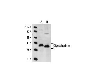
- Glycophorin A Anticorpo (R10) é recomendado para a detecção de Glycophorin A of human origin by WB, IP, IF, IHC(P) and FCM
- Anticorpo anti-Glycophorin A O anticorpo anti-Glycophorin A (R10) está disponível conjugado com agarose para IP; HRP para WB, IHC(P) e ELISA; e com fioeritrina ou FITC para IF, IHC(P) e FCM
- também disponível conjugado com Alexa Fluor® 488, Alexa Fluor® 546, Alexa Fluor® 594 ou Alexa Fluor® 647 para WB (RGB), IF, IHC(P) e FCM, e para utilização com sistemas de imagiologia fluorescente RGB, tais como iBright™ FL1000, FluorChem™, Typhoon, Azure e outros sistemas comparáveis
- também disponível conjugado com Alexa Fluor® 680 ou Alexa Fluor® 790 para WB (NIR), IF e FCM; para utilização com sistemas de deteção de infravermelhos próximos (NIR), tais como LI-COR®Odyssey®, iBright™ FL1000, FluorChem™, Typhoon, Azure e outros sistemas comparáveis
- m-IgG Fc BP-HRP, 1 BP-HRP">m-IgG1 BP-HRP e m-IgGκ BP-HRP são os reagentes de deteção secundários preferidos para Glycophorin A Antibody (R10) para aplicações WB e IHC(P). Estes reagentes são agora oferecidos em pacotes com Glycophorin A Antibody (R10)(ver informações de encomenda abaixo).

LINKS RÁPIDOS
VEJA TAMBÉM
O anticorpo anti-glicoforina A (R10) é um anticorpo monoclonal de cadeia leve IgG1 kappa de ratinho que detecta a glicoforina A de origem humana por western blotting (WB), imunoprecipitação (IP), imunofluorescência (IF), imunohistoquímica com secções incluídas em parafina (IHCP) e citometria de fluxo (FCM). O anticorpo anti-glicoforina A (R10) está disponível em formas não conjugadas e em várias formas conjugadas, incluindo agarose, peroxidase de rábano (HRP), ficoeritrina (PE), isotiocianato de fluoresceína (FITC) e vários conjugados Alexa Fluor®. A glicoforina A é uma sialoglicoproteína crucial localizada na superfície dos eritrócitos humanos, desempenhando um papel significativo na manutenção da integridade estrutural dos glóbulos vermelhos e facilitando a interação com o sistema imunitário. A glicoforina A atravessa a membrana uma vez, apresentando uma extremidade amino-terminal ao ambiente extracelular, essencial para a função antigénica do grupo sanguíneo. A diversidade genética dos antigénios de superfície da glicoforina, incluindo a glicoforina A, B e C, determina o fenótipo do grupo sanguíneo, tornando estas proteínas vitais para a compatibilidade da transfusão sanguínea e para a compreensão das doenças hemolíticas. O gene da Glicoforina A, localizado no cromossoma 4q31.21, é constituído por sete exões e apresenta uma elevada homologia com a Glicoforina B, codificando uma proteína de 91 aminoácidos. A compreensão da estrutura e da função da Glicoforina A continua a ser crucial para as aplicações clínicas e para a investigação da biologia dos eritrócitos, fornecendo informações sobre a expressão de antigénios de grupos sanguíneos e potenciais alvos terapêuticos para doenças relacionadas com o sangue.
Alexa Fluor® é uma marca comercial da Molecular Probes Inc., OR., EUA
LI-COR® e Odyssey® são marcas registadas da LI-COR Biosciences
Referencias do Glycophorin A Anticorpo (R10):
- Identificação imunohistoquímica de precursores eritróides em secções de medula óssea incluídas em parafina: a espectrina é um marcador superior à glicocoroína. | Sadahira, Y., et al. 1999. J Clin Pathol. 52: 919-21. PMID: 10711257
- Deteção in vivo da hetero-associação da glicoforina A e dos seus mutantes na membrana. | Gerber, D. and Shai, Y. 2001. J Biol Chem. 276: 31229-32. PMID: 11402026
- Regiões distintas da glicoforina A humana melhoram a função de transporte e o tráfico de superfície do permutador de aniões dos glóbulos vermelhos humanos (banda 3; AE1). | Young, MT. and Tanner, MJ. 2003. J Biol Chem. 278: 32954-61. PMID: 12813056
- Requisito da glicoforina A para a expressão de antigénios ligados a O na membrana eritrocitária. | Arimitsu, N., et al. 2003. Genes Cells. 8: 769-77. PMID: 12940824
- Alteração da estrutura e das propriedades de transporte de aniões da banda 3 (AE1, SLC4A1) em glóbulos vermelhos humanos com falta de glicoforina A. | Bruce, LJ., et al. 2004. J Biol Chem. 279: 2414-20. PMID: 14604989
- Adaptação estrutural do homodímero transmembranar da glicoforina A às modificações de D-aminoácidos. | Gerber, D., et al. 2004. J Mol Biol. 339: 243-50. PMID: 15123435
- Interacções complexas na interface hélice-hélice estabilizam o dímero transmembranar da glicoforina A. | Doura, AK. and Fleming, KG. 2004. J Mol Biol. 343: 1487-97. PMID: 15491626
- A co-transfecção de NXPE2 murino e de glicoforina A murina confere reatividade com Ter-119. | Keele, GR., et al. 2024. Haematologica. 109: 3755-3759. PMID: 39021224
- Glicoforina A como marcador de superfície celular da diferenciação eritroide precoce na leucemia aguda. | Andersson, LC., et al. 1979. Int J Cancer. 24: 717-20. PMID: 397196
- Expressão da glicoforina A na hematopoiese maligna. | Liszka, K., et al. 1983. Am J Hematol. 15: 219-26. PMID: 6638008
- Expressão de antigénios de superfície celular em progenitores eritróides humanos: marcadores eritróides e megacariocíticos. | Nakahata, T. and Okumura, N. 1994. Leuk Lymphoma. 13: 401-9. PMID: 8069185
- Diferenciação entre nódoas negras e descolorações putrefactivas da pele através da análise imunológica da glicoforina A. | Kibayashi, K., et al. 1993. Forensic Sci Int. 61: 111-7. PMID: 8307520
Informacoes sobre ordens
| Nome do Produto | Numero de Catalogo | UNID | Preco | Qde | FAVORITOS | |
Glycophorin A Anticorpo (R10) | sc-53905 | 200 µg/ml | $322.00 | |||
Pacote do Glycophorin A (R10): m-IgG Fc BP-HRP | sc-528587 | 200 µg Ab; 10 µg BP | $361.00 | |||
Pacote do Glycophorin A (R10): m-IgGκ BP-HRP | sc-520999 | 200 µg Ab, 40 µg BP | $361.00 | |||
Pacote do Glycophorin A (R10): m-IgG1 BP-HRP | sc-543011 | 200 µg Ab; 20 µg BP | $361.00 | |||
Glycophorin A Anticorpo (R10) AC | sc-53905 AC | 500 µg/ml, 25% agarose | $424.00 | |||
Glycophorin A Anticorpo (R10) HRP | sc-53905 HRP | 200 µg/ml | $322.00 | |||
Glycophorin A Anticorpo (R10) FITC | sc-53905 FITC | 200 µg/ml | $336.00 | |||
Glycophorin A Anticorpo (R10) PE | sc-53905 PE | 200 µg/ml | $349.00 | |||
Glycophorin A Anticorpo (R10) Alexa Fluor® 488 | sc-53905 AF488 | 200 µg/ml | $364.00 | |||
Glycophorin A Anticorpo (R10) Alexa Fluor® 546 | sc-53905 AF546 | 200 µg/ml | $364.00 | |||
Glycophorin A Anticorpo (R10) Alexa Fluor® 594 | sc-53905 AF594 | 200 µg/ml | $364.00 | |||
Glycophorin A Anticorpo (R10) Alexa Fluor® 647 | sc-53905 AF647 | 200 µg/ml | $364.00 | |||
Glycophorin A Anticorpo (R10) Alexa Fluor® 680 | sc-53905 AF680 | 200 µg/ml | $364.00 | |||
Glycophorin A Anticorpo (R10) Alexa Fluor® 790 | sc-53905 AF790 | 200 µg/ml | $364.00 |