")
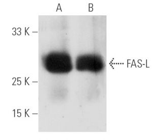
FAS-L Anticuerpo (NOK-1): sc-19681
- FAS-L Anticuerpo NOK-1 es un monoclonal de ratón IgG1 κ FAS-L Anticuerpo, ver las 37 publicaciones, proporcionado como 200 µg/ml
- producido contra células de ratón de T linfoma L5178Y expresando recombinante human FAS-L
- FAS-L Anticuerpo (NOK-1) es recomendado para detectar FAS-L de mouse, rat y human origen, mediante WB, IP, IF, IHC(P) y FCM; no recomendado para WB
- FAS-L Anticuerpo (NOK-1) es disponible conjugado a agarosa para IP; HRP para WB, IHC(P) y ELISA; y tanto a phycoerythrin como a FITC para IF, IHC(P) y FCM
- también disponible conjugado a Alexa Fluor® 488, Alexa Fluor® 546, Alexa Fluor® 594 o Alexa Fluor® 647 para WB (RGB), IF, IHC (P) y FCM
- también disponible conjugado a Alexa Fluor® 680 o Alexa Fluor® 790 para WB (NIR), IF y FCM
- también se suministra sin azida para neutralizar los efectos citotóxicos de FAS-L, sc-19681 L, 200 µg/0,1 ml
- m-IgG Fc BP-HRP y m-IgG1 BP-HRP son los reactivos de detección secundarios preferidos para FAS-L Anticuerpo (NOK-1) for WB and IHC(P) applications. Estos reactivos se ofrecen ahora en paquetes con FAS-L Anticuerpo (NOK-1)(véase la información de pedido más abajo).

ENLACES RÁPIDOS
VER TAMBIÉN ....
El anticuerpo FAS-L (NOK-1) es un anticuerpo monoclonal IgG1 de ratón de cadena ligera kappa que detecta la proteína FAS-L (también conocida como FASLG, CD178, CD95-L, TNFSF6 o ligando de Fas) en muestras de ratón, rata y humanos mediante western blotting, inmunoprecipitación, inmunofluorescencia, inmunohistoquímica en secciones embebidas en parafina y citometría de flujo. El anticuerpo FAS-L (NOK-1) está disponible en forma no conjugada y en múltiples formas conjugadas, incluyendo agarosa, peroxidasa de rábano picante, ficoeritrina, isotiocianato de fluoresceína y múltiples conjugados Alexa Fluor®. La citotoxicidad mediada por linfocitos T citotóxicos (CTL) constituye un componente importante de los mecanismos efectores específicos en la inmunovigilancia contra células infectadas o transformadas por virus. Dos mecanismos explican esta actividad, uno de los cuales es el proceso basado en la perforina. Un mecanismo basado en FAS implica a la molécula transductora FAS (también conocida como Apo-1) y FAS-L. La proteína FAS humana es una glicoproteína de la superficie celular que pertenece a una familia de receptores que incluye el CD40, los receptores del factor de crecimiento nervioso y los receptores del factor de necrosis tumoral. El antígeno FAS se expresa en una amplia gama de líneas celulares linfoides, algunas de las cuales sufren apoptosis en respuesta al tratamiento con anticuerpo anti-FAS. Estos hallazgos implican fuertemente que la muerte celular dirigida está potencialmente mediada por interacciones intercelulares de FAS con FAS-L o efectores, y que FAS puede estar críticamente implicado en la citotoxicidad mediada por CTL.
Alexa Fluor® es una marca registrada de Molecular Probes Inc., OR., USA
REIVEW LI-COR® y Odyssey® son marcas registradas de LI-COR Biosciences.
FAS-L Anticuerpo (NOK-1) Referencias:
- Modulación del fas-ligando (Fas-L) en las células microgliales humanas: un estudio in vitro. | Frigerio, S., et al. 2000. J Neuroimmunol. 105: 109-14. PMID: 10742551
- Mecanismo de escape tumoral que implica a las moléculas Fas y Fas-L en los tumores colorrectales humanos. | Radfar, S., et al. 2000. Gastroenterol Clin Biol. 24: 1191-6. PMID: 11173732
- Regulación al alza de Fas-L por células plasmáticas de mieloma altamente maligno: papel en la patogénesis de la anemia y la progresión de la enfermedad. | Silvestris, F., et al. 2001. Blood. 97: 1155-64. PMID: 11222356
- El papel crítico de la proteína quinasa C-theta en la apoptosis mediada por el ligando Fas/Fas. | Manicassamy, S. and Sun, Z. 2007. J Immunol. 178: 312-9. PMID: 17182568
- FasL y TRAIL inducen la apoptosis epidérmica y la ulceración cutánea tras la exposición a Leishmania major. | Eidsmo, L., et al. 2007. Am J Pathol. 170: 227-39. PMID: 17200196
- [Expresión de gastrina, somatostatina, PCNA y Fas-L en la mucosa del antro gástrico de niños con gastritis crónica y úlcera duodenal]. | Xie, XZ., et al. 2006. Zhonghua Er Ke Za Zhi. 44: 774-7. PMID: 17229384
- Los mediadores de la apoptosis Fas y FasL predicen la progresión de la discapacidad en la esclerosis múltiple durante un periodo de 10 años. | Lopatinskaya, L., et al. 2006. Mult Scler. 12: 704-9. PMID: 17262997
- La eliminación de células T reguladoras CD4+CD25+ intratumorales mediante la transferencia de la proteína FasL mejora la eficacia terapéutica de la transferencia adoptiva de células T. | Chen, A., et al. 2007. Cancer Res. 67: 1291-8. PMID: 17283166
- [La apoptosis de los linfocitos alveolares en la sarcoidosis y en el grupo de control es más frecuente en los fumadores que en los no fumadores]. | Kopiński, P., et al. 2006. Przegl Lek. 63: 841-7. PMID: 17288168
- Mecanismo de la citotoxicidad mediada por linfocitos. | Henkart, PA. 1985. Annu Rev Immunol. 3: 31-58. PMID: 3904772
- Fas y su ligando en un mecanismo general de citotoxicidad mediada por células T. | Hanabuchi, S., et al. 1994. Proc Natl Acad Sci U S A. 91: 4930-4. PMID: 7515183
- La proteína Fas se expresa a niveles elevados en timocitos CD4+CD8+ y linfocitos maduros activados en ratones normales, pero no en la cepa propensa al lupus, MRL lpr/lpr. | Drappa, J., et al. 1993. Proc Natl Acad Sci U S A. 90: 10340-4. PMID: 7694292
Información sobre pedidos
| Nombre del producto | Número de catálogo | UNIDAD | Precio | CANTIDAD | Favoritos | |
FAS-L Anticuerpo (NOK-1) | sc-19681 | 200 µg/ml | $322.00 | |||
Paquete de FAS-L (NOK-1): m-IgG Fc BP-HRP | sc-526589 | 200 µg Ab; 10 µg BP | $361.00 | |||
Paquete de FAS-L (NOK-1): m-IgG1 BP-HRP | sc-531962 | 200 µg Ab; 20 µg BP | $361.00 | |||
FAS-L Anticuerpo (NOK-1) L | sc-19681 L | 200 µg/0.1 ml | $322.00 | |||
FAS-L Anticuerpo (NOK-1) AC | sc-19681 AC | 500 µg/ml, 25% agarose | $424.00 | |||
FAS-L Anticuerpo (NOK-1) HRP | sc-19681 HRP | 200 µg/ml | $322.00 | |||
FAS-L Anticuerpo (NOK-1) FITC | sc-19681 FITC | 200 µg/ml | $336.00 | |||
FAS-L Anticuerpo (NOK-1) PE | sc-19681 PE | 200 µg/ml | $349.00 | |||
FAS-L Anticuerpo (NOK-1) Alexa Fluor® 488 | sc-19681 AF488 | 200 µg/ml | $364.00 | |||
FAS-L Anticuerpo (NOK-1) Alexa Fluor® 546 | sc-19681 AF546 | 200 µg/ml | $364.00 | |||
FAS-L Anticuerpo (NOK-1) Alexa Fluor® 594 | sc-19681 AF594 | 200 µg/ml | $364.00 | |||
FAS-L Anticuerpo (NOK-1) Alexa Fluor® 647 | sc-19681 AF647 | 200 µg/ml | $364.00 | |||
FAS-L Anticuerpo (NOK-1) Alexa Fluor® 680 | sc-19681 AF680 | 200 µg/ml | $364.00 | |||
FAS-L Anticuerpo (NOK-1) Alexa Fluor® 790 | sc-19681 AF790 | 200 µg/ml | $364.00 |