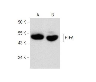
Date published: 2025-9-7

00800 4573 8000

SCBT Portrait Logo
Seach Input

ETEA Antikörper (F-7): sc-374098

4.7(3)
Produkt bewertenBitte stellen Sie eine Frage

Datenblätter
  • ETEA Antikörper F-7 ist ein Maus monoklonales IgG1 κ ETEA Antikörper, verwendet in 4 wissenschaftlichen Veröffentlichungen, in einer Menge von 200 µg/ml
  • gezogen gegen die Aminosäuresequenz 147-434 lokalisiert am C-terminus von ETEA aus der Spezies human
  • ETEA Antikörper (F-7) ist empfohlen für die Detektion von ETEA aus der Spezies mouse, rat und human per WB, IP, IF und ELISA
  • Anti-ETEA Antikörper (F-7) ist erhältlich als Konjugat mit Agarose für IP; HRP für WB, IHC(P) und ELISA; und entweder mit Phycoerythrin oder FITC für IF, IHC(P) und FCM
  • auch erhältlich als Konjugat mit Alexa Fluor® 488, Alexa Fluor® 546, Alexa Fluor® 594 oder Alexa Fluor® 647 für IF, IHC(P) und FCM
  • auch erhältlich als Konjugat mit Alexa Fluor® 680 oder Alexa Fluor® 790 für WB (NIR), IF und FCM
  • Bitte kontaktieren Sie uns um ein 10 µg GRATIS Sample vom ETEA (F-7): sc-374098 zu erhalten.
  • m-IgG Fc BP-HRP und 1 BP-HRP">m-IgG1 BP-HRP sind die bevorzugten sekundären Nachweisreagenzien für ETEA Antikörper (F-7) for WB applications.. Diese Reagenzien werden jetzt in Bündeln mit ETEA Antikörper (F-7) angeboten(siehe Bestellinformationen unten).

Direktverknüpfungen

Siehe auch...

Der ETEA-Antikörper (F-7) ist ein monoklonaler Maus IgG1 κ ETEA-Antikörper (auch als ETEA-Antikörper bezeichnet), der das ETEA-Protein von Maus-, Ratte- und menschlicher Herkunft mittels WB, IP, IF und ELISA nachweist. Der ETEA-Antikörper (F-7) ist sowohl in nicht konjugierter Form als auch in mehreren konjugierten Formen des ETEA-Antikörpers erhältlich, darunter Agarose, HRP, PE, FITC und mehrere Alexa Fluor®-Konjugate. ETEA, auch als UBX-Domänen-Protein 8 (UBXD8) bezeichnet, ist ein 445-Aminosäure-Protein, das mit atopischer Dermatitis (AD) in Verbindung gebracht wird, einer chronischen, nicht ansteckenden, rezidivierenden entzündlichen Hauterkrankung, die durch ekzematöse Hautläsionen gekennzeichnet ist und auch als ekzematöse Dermatitis bezeichnet wird. Andere atopische Erkrankungen wie Heuschnupfen, Asthma und Konjunktivitis treten häufig zusammen mit AD auf. ETEA zeigt in T-Zellen und Eosinophilen von Patienten mit AD eine höhere Expression als in T-Zellen und Eosinophilen unbetroffener Personen. T-Zellen spielen bei der Regulation des Entzündungsprozesses dieser Erkrankung eine wichtige Rolle. Die Persistenz von jAD wird auf eine dysregulierte Apoptose in T-Zellen, Eosinophilen und Keratinozyten zurückgeführt. ETEA könnte an der Resistenz gegen Apoptose in T-Zellen und Eosinophilen von AD-Patienten beteiligt sein.

Ausschließlich für Forschungszwecke. Nicht für diagnostische oder therapeutische Zwecke bestimmt.

Alexa Fluor® ist ein Markenzeichen von Molecular Probes Inc., OR., USA

LI-COR® und Odyssey® sind Markenzeichen von LI-COR Biosciences

ETEA Antikörper (F-7) Literaturhinweise:

  1. Atopische Dermatitis: neue Erkenntnisse und Möglichkeiten für therapeutische Maßnahmen.  |  Leung, DY. 2000. J Allergy Clin Immunol. 105: 860-76. PMID: 10808164
  2. T-Zell-vermittelte Fas-induzierte Keratinozyten-Apoptose spielt eine wichtige pathogenetische Rolle bei ekzematöser Dermatitis.  |  Trautmann, A., et al. 2000. J Clin Invest. 106: 25-35. PMID: 10880045
  3. Die Rolle der dysregulierten Apoptose bei atopischer Dermatitis.  |  Trautmann, A., et al. 2000. Apoptosis. 5: 425-9. PMID: 11256884
  4. Die Rolle der Apoptose bei atopischer Dermatitis.  |  Trautmann, A., et al. 2001. Int Arch Allergy Immunol. 124: 230-2. PMID: 11306978
  5. Klonierung und Charakterisierung des hochexprimierten ETEA-Gens aus Blutzellen von Patienten mit atopischer Dermatitis.  |  Imai, Y., et al. 2002. Biochem Biophys Res Commun. 297: 1282-90. PMID: 12372427
  6. [Lymphozyten-Apoptose bei atopischer Dermatitis, die mit Transferfaktor behandelt wird].  |  García Angeles, J., et al. 2003. Rev Alerg Mex. 50: 3-7. PMID: 12822541

Bestellinformation

ProduktKatalog #EINHEITPreisANZAHLFavoriten

ETEA Antikörper (F-7)

sc-374098
200 µg/ml
$316.00

ETEA (F-7): m-IgG Fc BP-HRP Bundle

sc-527434
200 µg Ab; 10 µg BP
$354.00

ETEA (F-7): m-IgG1 BP-HRP Bundle

sc-532807
200 µg Ab; 20 µg BP
$354.00

ETEA Antikörper (F-7) AC

sc-374098 AC
500 µg/ml, 25% agarose
$416.00

ETEA Antikörper (F-7) HRP

sc-374098 HRP
200 µg/ml
$316.00

ETEA Antikörper (F-7) FITC

sc-374098 FITC
200 µg/ml
$330.00

ETEA Antikörper (F-7) PE

sc-374098 PE
200 µg/ml
$343.00

ETEA Antikörper (F-7) Alexa Fluor® 488

sc-374098 AF488
200 µg/ml
$357.00

ETEA Antikörper (F-7) Alexa Fluor® 546

sc-374098 AF546
200 µg/ml
$357.00

ETEA Antikörper (F-7) Alexa Fluor® 594

sc-374098 AF594
200 µg/ml
$357.00

ETEA Antikörper (F-7) Alexa Fluor® 647

sc-374098 AF647
200 µg/ml
$357.00

ETEA Antikörper (F-7) Alexa Fluor® 680

sc-374098 AF680
200 µg/ml
$357.00

ETEA Antikörper (F-7) Alexa Fluor® 790

sc-374098 AF790
200 µg/ml
$357.00

What is the recommended fixation method for immunofluorescence staining with ETEA (F-7): sc-374098 monoclonal antibody?

Gefragt von: DefinitelyNotMatt
Thank you for your inquiry. We recommend fixing cells for 5 minutes in -10°C methanol and allowing to air dry. The full protocol can be viewed here: https://www.scbt.com/scbt/resources/protocols/immunofluorescence-cell-staining
Beantwortet von: Technical Support
Veröffentlichungsdatum: 2017-02-24
  • y_2025, m_9, d_7, h_4CST
  • bvseo_bulk, prod_bvqa, vn_bulk_3.0.42
  • cp_1, bvpage1
  • co_hasquestionsanswers, tq_1
  • loc_de_DE, sid_374098, prod, sort_[SortEntry(order=LAST_APPROVED_ANSWER_SUBMISSION_TIME, direction=DESCENDING)]
  • clientName_scbt
  • bvseo_sdk, java_sdk, bvseo-4.0.0
  • CLOUD, getContent, 101ms
  • QUESTIONS, PRODUCT
Rated 4 von 5 von aus Good antibody for WB in HeLa cell lysateGood antibody for WB in HeLa cell lysate.
Veröffentlichungsdatum: 2015-12-14
Rated 5 von 5 von aus Produced superb immunofluorescence cytoplasmicProduced superb immunofluorescence cytoplasmic staining in methanol-fixed HeLa cells. -SCBT QC
Veröffentlichungsdatum: 2015-05-14
Rated 5 von 5 von aus Nice Western Blot data of ETEA expressionNice Western Blot data of ETEA expression in Jurkat and MOLT-4 whole cell lysates. -SCBT QC
Veröffentlichungsdatum: 2013-11-25
  • y_2025, m_9, d_7, h_4
  • bvseo_bulk, prod_bvrr, vn_bulk_3.0.42
  • cp_1, bvpage1
  • co_hasreviews, tv_0, tr_3
  • loc_de_DE, sid_374098, prod, sort_[SortEntry(order=SUBMISSION_TIME, direction=DESCENDING)]
  • clientName_scbt
  • bvseo_sdk, java_sdk, bvseo-4.0.0
  • CLOUD, getReviews, 17ms
  • REVIEWS, PRODUCT
ETEA Antikörper (F-7) wurde bewertet mit 4.7 von 5 von 3.
  • y_2025, m_9, d_7, h_4
  • bvseo_bulk, prod_bvrr, vn_bulk_3.0.42
  • cp_1, bvpage1
  • co_hasreviews, tv_0, tr_3
  • loc_de_DE, sid_374098, prod, sort_[SortEntry(order=SUBMISSION_TIME, direction=DESCENDING)]
  • clientName_scbt
  • bvseo_sdk, java_sdk, bvseo-4.0.0
  • CLOUD, getAggregateRating, 107ms
  • REVIEWS, PRODUCT