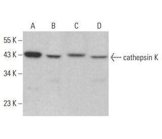
Date published: 2025-9-7

00800 4573 8000

SCBT Portrait Logo
Seach Input

cathepsin K Antikörper (E-7): sc-48353

5.0(20)
Produkt bewertenBitte stellen Sie eine Frage

Datenblätter
  • cathepsin K Antikörper E-7 ist ein Maus monoklonales IgG1 κ cathepsin K Antikörper, verwendet in 165 wissenschaftlichen Veröffentlichungen, in einer Menge von 200 µg/ml
  • gegen Aminosäuren 191-240 gerichtet, die in einer internen Region von cathepsin K mit Ursprung human kartiert werden
  • Empfohlen für die Detektion von cathepsin K aus der Spezies mouse, rat und human per WB, IP, IF, IHC(P) und ELISA
  • erhältlich als Konjugat mit HRP für WB, IHC(P) und ELISA; und entweder mit Phycoerythrin oder FITC, Alexa Fluor® 488 oder Alexa Fluor® 647 für IF, IHC(P) und FCM
  • Bitte kontaktieren Sie uns um ein 10 µg GRATIS Sample vom cathepsin K (E-7): sc-48353 zu erhalten.
  • m-IgG Fc BP-HRP und m-IgGκ BP-HRP sind die empfohlenen Detektionsreagenzien für cathepsin K Antikörper (E-7) für WB and IHC(P) Experimente. Diese Reagenzien werden nun auch als Bundle angeboten, zusammen mit cathepsin K Antikörper (E-7) (Bestellinformation unten stehend).

Direktverknüpfungen

Siehe auch...

Der Cathepsin-K-Antikörper (E-7) ist ein monoklonaler IgG1-Antikörper der leichten Kette von Mäusen, der das Cathepsin-K-Protein von Mäusen, Ratten und Menschen durch Western Blot (WB), Immunopräzipitation (IP), Immunfluoreszenz (IF), Immunhistochemie mit in Paraffin eingebetteten Schnitten (IHCP) und Enzyme-linked Immunosorbent Assay (ELISA) nachweist. Der Anti-Cathepsin-K-Antikörper (E-7) ist sowohl in nicht konjugierter als auch in verschiedenen konjugierten Formen erhältlich, darunter Agarose, Meerrettichperoxidase (HRP), Phycoerythrin (PE), Fluoresceinisothiocyanat (FITC) und mehrere Alexa Fluor®-Konjugate. Cathepsin K, ein Mitglied der Cysteinprotease-Familie, spielt eine entscheidende Rolle bei der Knochenresorption und -remodellierung, wodurch Cathepsin K für die Aufrechterhaltung der Knochengesundheit und -homöostase unerlässlich ist. Dieses Enzym wird vorwiegend in Osteoklasten exprimiert, Zellen, die für den Knochenabbau verantwortlich sind, und die Cathepsin-K-Aktivität ist für einen normalen physiologischen Knochenumsatz unerlässlich. Eine Dysregulation von Cathepsin K wurde mit verschiedenen Knochenerkrankungen in Verbindung gebracht, darunter Osteoporose und Morbus Paget, was die Bedeutung von Cathepsin K als therapeutisches Ziel unterstreicht. Die Fähigkeit des Anti-Cathepsin-K-Antikörpers (E-7), mehrere Spezies zu erkennen, verbessert die Forschung und klinische Anwendungen und ermöglicht ein tieferes Verständnis des Knochenstoffwechsels und damit zusammenhängender Krankheiten.

Ausschließlich für Forschungszwecke. Nicht für diagnostische oder therapeutische Zwecke bestimmt.

Alexa Fluor® ist ein Markenzeichen von Molecular Probes Inc., OR., USA

LI-COR® und Odyssey® sind Markenzeichen von LI-COR Biosciences

cathepsin K Antikörper (E-7) Literaturhinweise:

  1. Molekulare Klonierung und Expression von menschlichem alveolärem Makrophagen-Cathepsin S, einer elastinolytischen Cysteinprotease.  |  Shi, GP., et al. 1992. J Biol Chem. 267: 7258-62. PMID: 1373132
  2. Molekulare Organisation des menschlichen Cathepsin-D-Gens.  |  Redecker, B., et al. 1991. DNA Cell Biol. 10: 423-31. PMID: 2069717
  3. Cathepsin-K-Inhibitoren zur Behandlung von Osteoporose: Biologie, potenzieller klinischer Nutzen und Lehren aus den Erfahrungen.  |  Drake, MT., et al. 2017. Endocr Rev. 38: 325-350. PMID: 28651365
  4. Cathepsin K-exprimierende Vorläuferzellen aus der Sehne aktivieren die Hedgehog-Signalübertragung, um die heterotope Verknöcherung voranzutreiben.  |  Feng, H., et al. 2020. J Clin Invest. 130: 6354-6365. PMID: 32853181
  5. Molekulare Klonierung und Sequenzierung der cDNA für Ratten-Cathepsin L.  |  Ishidoh, K., et al. 1987. FEBS Lett. 223: 69-73. PMID: 3666143
  6. Molekulare Klonierung und Sequenzierung der cDNA für Ratten-Cathepsin H. Homologie in Pro-Peptid-Regionen von Cysteinproteinasen.  |  Ishidoh, K., et al. 1987. FEBS Lett. 226: 33-7. PMID: 3691815
  7. Die Hemmung von Cathepsin K führt über die Syk/SHP2/Src/OTUB1-Achse zu einer Destabilisierung von Raptor und einer mitochondrialen Dysfunktion.  |  Seo, SU., et al. 2023. Cell Death Dis. 14: 366. PMID: 37330581
  8. Isolierung und Sequenzierung von zwei cDNA-Klonen, die für Rattenmilz-Cathepsin E kodieren, und Analyse der Aktivierung von gereinigtem Procathepsin E.  |  Okamoto, K., et al. 1995. Arch Biochem Biophys. 322: 103-11. PMID: 7574663
  9. Molekulare Klonierung von menschlichem Cathepsin O, einer neuen Endoproteinase und Homolog von Kaninchen OC2.  |  Shi, GP., et al. 1995. FEBS Lett. 357: 129-34. PMID: 7805878
  10. Cathepsin B, eine Cysteinprotease, die bei der Metastasenbildung eine Rolle spielt, wird auch bei der Rückbildung der Rattenprostata und der Brustdrüsen exprimiert.  |  Guenette, RS., et al. 1994. Eur J Biochem. 226: 311-21. PMID: 8001549
  11. Molekulare Klonierung, chromosomale Lage und gewebespezifische Expression des murinen Cathepsin-G-Gens.  |  Heusel, JW., et al. 1993. Blood. 81: 1614-23. PMID: 8453108
  12. Maus-Cathepsin K: cDNA-Klonierung und vorherrschende Expression des Gens in Osteoklasten und in einigen hypertrophierenden Chondrozyten während der Mausentwicklung.  |  Rantakokko, J., et al. 1996. FEBS Lett. 393: 307-13. PMID: 8814310

Bestellinformation

ProduktKatalog #EINHEITPreisANZAHLFavoriten

cathepsin K Antikörper (E-7)

sc-48353
200 µg/ml
$316.00

cathepsin K (E-7): m-IgG Fc BP-HRP Bundle

sc-528526
200 µg Ab; 10 µg BP
$354.00

cathepsin K (E-7): m-IgGκ BP-HRP Bundle

sc-520922
200 µg Ab, 40 µg BP
$354.00

cathepsin K Antikörper (E-7) Alexa Fluor® 488

sc-48353 AF488
200 µg/ml
$357.00

cathepsin K Antikörper (E-7) Alexa Fluor® 647

sc-48353 AF647
200 µg/ml
$357.00

cathepsin K Antikörper (E-7) FITC

sc-48353 FITC
200 µg/ml
$330.00

cathepsin K Antikörper (E-7) HRP

sc-48353 HRP
200 µg/ml
$316.00

cathepsin K Antikörper (E-7) PE

sc-48353 PE
200 µg/ml
$343.00

I cannot find an antibody to Cathepsin K that can be used in rabbit. There are Cathepsin K antibodies available raised in rabbit but that should give a very high back ground and may lead to false positives. Any suggestions?

Gefragt von: Doty
Thank you for your question, this antibody is not warrantied for rabbit, but it does have a high homology for rabbit. This would be what we'd recommend for your sample.
Beantwortet von: Technical Service 4
Veröffentlichungsdatum: 2020-12-21

What concentration should this be used at for flow cytometry? Presumably this is after fix/perm?

Gefragt von: SB KCL
Thank you for your question. Please note that unconjugated cathepsin K Antibody (E-7): sc-48353 is not recommended for flow cytometry. You can find our standard protocol under this link: https://www.scbt.com/resources/protocols/flow-cytometry We recommend using 1 µg of fluorochrome-conjugated antibodies per tube with 1 million cells.
Beantwortet von: Technical Support Europe
Veröffentlichungsdatum: 2020-10-21

For WB, is it possible to use secondary Fc-specific antibody to detect E-7 (sc-48353) antibody ?

Gefragt von: TomG
Thank you for your inquiry and your interest in this product. The cathepsin K Antibody (E-7): sc-48353 is a mouse monoclonal IgG3 containing a kappa light chain. You can certainly use a conventional Fc-specific anti-mouse secondary antibody for detection. However, we would rather recommend to use the directly conjugated formats of this antibody or our Mouse IgG Kappa Binding Proteins, e.g. m-IgGκ BP-HRP: sc-516102. Please contact our European Support Office in Germany directly for any further questions you may have. You can reach us by phone at: +49 6221 4503 0, by email at: europe@scbt.com or by live chat directly on our website, www.scbt.com We are happy to assist you.
Beantwortet von: Technical Support Europe
Veröffentlichungsdatum: 2018-01-04

For IHC, which antigen retrieval method is recommended?

Gefragt von: rbrun
Thank you for your question. We recommend using sodium citrate buffer (pH 6.0) with heat treatment for antigen retrieval with this antibody, cathepsin K (E-7): sc-48353. A link to our recommended protocol is provided below as a reference. https://www.scbt.com/scbt/resources/protocols/immunoperoxidase-staining
Beantwortet von: Technical Support
Veröffentlichungsdatum: 2024-12-03

For Western Blot, is it recommended to use denatured or non-denatured conditions with this antibody?

Gefragt von: AbPolly
Thank you for your question. We recommend this antibody for use in denatured Western Blot conditions. It has not been validated for use in non-denatured conditions. Please contact our Technical Service Department for further details or inquiries.
Beantwortet von: Technical Support 12
Veröffentlichungsdatum: 2017-01-04
  • y_2025, m_8, d_28, h_5CST
  • bvseo_bulk, prod_bvqa, vn_bulk_3.0.42
  • cp_1, bvpage1
  • co_hasquestionsanswers, tq_5
  • loc_de_DE, sid_48353, prod, sort_[SortEntry(order=LAST_APPROVED_ANSWER_SUBMISSION_TIME, direction=DESCENDING)]
  • clientName_scbt
  • bvseo_sdk, java_sdk, bvseo-4.0.0
  • CLOUD, getContent, 105ms
  • QUESTIONS, PRODUCT
Rated 5 von 5 von aus 非常好的一只抗体!在Western blot实验中表现出色一年多前购买了这只抗体,效果非常的不错。非常好的一只抗体!在Western blot实验中表现出色!
Veröffentlichungsdatum: 2024-12-02
Rated 5 von 5 von aus Very specific antibodyWe used these antibody to analyze active cathepsin K in mouse and human osteoclast. You can see our results in the following publication: Dey, G. et al, Development and Application of Reversible and Irreversible Covalent Probes for Human and Mouse Cathepsin‐K Activity Detection, Revealing Nuclear Activity. Advanced Science 2024. https://doi.org/10.1002/advs.202401518.
Veröffentlichungsdatum: 2024-11-25
Rated 4 von 5 von aus Cathepsin KDot Blot was perfomed to detect the Cathepsin K in the cell culture supernatants.
Veröffentlichungsdatum: 2020-07-29
Rated 5 von 5 von aus Great signal!Works well in 1:500 dilution to stain cathepsin K from mouse bone marrow-derived osteoclast samples.
Veröffentlichungsdatum: 2019-03-22
Rated 5 von 5 von aus Works well1:500 dilution, clear bands in mouse BMMs induced to osteoclast by RANKL
Veröffentlichungsdatum: 2018-01-17
Rated 5 von 5 von aus Excellent stainingI have received this sample and used it for staining of cat K in mice aortic tissue and cultured vascular smooth muscle cells. The staining was very clear and excellent.
Veröffentlichungsdatum: 2017-10-27
Rated 5 von 5 von aus great signalUsing 1/1000 diluted the antibody to work in WB, and there were bands in the mouse tissue, ~39kDa, which was a good result.
Veröffentlichungsdatum: 2017-10-24
Rated 5 von 5 von aus Worked very well!I tried IHC-P in mouse tissue (bone) and the results were very nice at 1:100.
Veröffentlichungsdatum: 2017-09-22
  • y_2025, m_8, d_28, h_5
  • bvseo_bulk, prod_bvrr, vn_bulk_3.0.42
  • cp_1, bvpage1
  • co_hasreviews, tv_0, tr_20
  • loc_de_DE, sid_48353, prod, sort_[SortEntry(order=SUBMISSION_TIME, direction=DESCENDING)]
  • clientName_scbt
  • bvseo_sdk, java_sdk, bvseo-4.0.0
  • CLOUD, getReviews, 19ms
  • REVIEWS, PRODUCT
cathepsin K Antikörper (E-7) wurde bewertet mit 5.0 von 5 von 20.
  • y_2025, m_8, d_28, h_5
  • bvseo_bulk, prod_bvrr, vn_bulk_3.0.42
  • cp_1, bvpage1
  • co_hasreviews, tv_0, tr_20
  • loc_de_DE, sid_48353, prod, sort_[SortEntry(order=SUBMISSION_TIME, direction=DESCENDING)]
  • clientName_scbt
  • bvseo_sdk, java_sdk, bvseo-4.0.0
  • CLOUD, getAggregateRating, 153ms
  • REVIEWS, PRODUCT