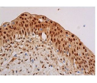
Date published: 2025-9-8

00800 4573 8000

SCBT Portrait Logo
Seach Input

p-Akt1/2/3 Anticorpo (B-12): sc-377556

4.2(10)
Escrever uma avaliaçãoFazer uma pergunta

Fichas de dados
  • p-Akt1/2/3 Anticorpo B-12é um anticorpo monoclonal produzido em camundongo IgM κ, citado em 38 publicações, fornecido em 200 µg/ml
  • específica para um mapeamento de epítopos entre os aminoácidos Thr 450 Thr 450 de Akt1 de human origem
  • recomendado para a detecção de Thr 450 phosphorylated Akt1 and Thr 451 correspondingly phosphorylated Akt2 and Thr 447 correspondingly phosphorylated Akt3 de mouse, rat e human origem em; Reage também com outras espécies, incluindo and equine, bovine and porcine
  • Entre em contato conosco e receba GRATUITAMENTE um amostra de 10 µg de p-Akt1/2/3 (B-12): sc-377556.
  • Atualmente, ainda não concluímos a identificação do(s) reagente(s) de deteção secundário(s) preferido(s) para p-Akt1/2/3 Anticorpo (B-12). Este trabalho está em curso.

LINKS RÁPIDOS

VEJA TAMBÉM

O anticorpo p-Akt1/2/3 (B-12) é um anticorpo monoclonal de camundongo IgM κ que detecta a Akt1 fosforilada na Treonina 450 e, correspondendo a isso, a Akt2 fosforilada na Treonina 451 e a Akt3 fosforilada na Treonina 447, de origem de camundongo, rato e humano, por WB, IP, IF, IHC(P) e ELISA. O anticorpo p-Akt1/2/3 (B-12) está disponível na forma de anticorpo anti-p-Akt1/2/3 não conjugado. A família de quinases serina/treonina Akt contém vários membros, incluindo Akt1 (também designado PKB ou RacPK), Akt2 (também designado PKBβ ou RacPK-β) e Akt 3 (também designado PKBγ ou proto-oncogene viral de timoma 3), que exibem homologia de sequência com as famílias de quinase de proteína A e C e são codificados pelo proto-oncogene c-Akt. Todos os membros da família Akt possuem um domínio de homologia de pleckstrina. Akt1 e Akt2 são ativados pela estimulação do PDGF. Essa ativação depende dos resíduos de tirosina 740 e 751 do PDGFR-β, que se ligam à subunidade do complexo de fosfatidilinositol 3-quinase (PI 3-quinase). A ativação de Akt1 pela insulina ou pelo fator de crescimento semelhante à insulina-1(IGF-1) resulta na fosforilação tanto de Treonina 308 quanto de Serina 473. As proteínas Akt tornam-se fosforiladas e ativadas em células estimuladas por insulina/IGF-1 por uma quinase(s) arduas, e a ativação de Akt1 e Akt2 é inibida pelo inibidor da quinase PI wortmannin. Em conjunto, esses dados sugerem que a proteína sinaliza a jusante das quinases PI. A Akt3 é fosforilada em um resíduo de serina em resposta à insulina. No entanto, a ativação da Akt3 pela insulina é inibida pela ativação prévia da proteína quinase C por um mecanismo que não requer a presença do domínio PH. A Akt3 é expressa em fibroblastos 3T3-L1, adipócitos e músculo esquelético e pode estar envolvida em vários processos biológicos, incluindo diferenciação de adipócitos e músculos, síntese de glicogênio, captação de glicose, apoptose e proliferação celular.

Para uso em exclusivo em pesquisa. Não se destina a uso em diagnostico e tratamento.

Informacoes sobre ordens

Nome do ProdutoNumero de CatalogoUNIDPrecoQdeFAVORITOS

p-Akt1/2/3 Anticorpo (B-12)

sc-377556
200 µg/ml
$316.00

p-Akt1/2/3 (B-12) peptídeo neutralizante

sc-377556 P
100 µg/0.5 ml
$68.00

I am using B-12 antibody (pAKT), is it igM second antibody with HRP to be used for detection? or will it work with igGk second antibody?

Asked by: Chai 123
Thank you for your question. p-Akt1/2/3 Antibody (B-12) is a mouse monoclonal IgM and we do recommend using an isotype specific secondary anti-mouse IgM for detection. Alternatively, you could also use our mouse IgG kappa binding protein (m-IgGκ BP) conjugated to Horseradish Peroxidase (HRP), m-IgGκ BP-HRP: sc-516102. This secondary antibody binds specifically to the kappa light chain of mouse IgG, and is also reactive with the kappa light chain of mouse IgM. A link to the product information is provided below for your reference. https://www.scbt.com/scbt/product/m-igg-kappa-bp-hrp
Answered by: Technical Support
Date published: 2017-06-29

What application is the blocking peptide sc-377556 P appropriate for?

Asked by: DefinitelyNotMatt
Thank you for your question. The blocking peptide is intended for use as a negative control, by pre-adsorbing the mouse monoclonal antibody against the antigen. For full protocol details, please contact our Technical Services Department or view our online protocol here: https://www.scbt.com/scbt/resources/protocols/peptide-neutralization
Answered by: Technical Support
Date published: 2017-02-24
  • y_2025, m_8, d_27, h_5CST
  • bvseo_bulk, prod_bvqa, vn_bulk_3.0.42
  • cp_1, bvpage1
  • co_hasquestionsanswers, tq_2
  • loc_pt_PT, sid_377556, prod, sort_[SortEntry(order=LAST_APPROVED_ANSWER_SUBMISSION_TIME, direction=DESCENDING)]
  • clientName_scbt
  • bvseo_sdk, java_sdk, bvseo-4.0.0
  • CLOUD, getContent, 104ms
  • QUESTIONS, PRODUCT
Rated 4 out of 5 by from Showed specific bandsGood antibody showed specific band with 1:500 dilution.
Date published: 2017-10-20
Rated 4 out of 5 by from Not very sureSample was mixed with 5X Reducing Loading Buffer and Denature the proteins by heating the sample to 95°C. Load the sample onto 3% Stacking Gel and 10% Separation Gel and run the gel under standard conditions. Transfer the proteins to a PVDF membrane using semi-dry or wet transfer methods. Block the membrane with 3% w/v skim milk powder in TBST. Incubate for 2 hr with agitation. Dilute the primary antibody in WB Anti-body Diluent (1:200) to the recommended dilution. Incubate overnight at 4°C, and rinse the blot in TBST four times for 5 min each at room temperature. Dilute the HRP labeled goat anti-mouse secondary antibody at the Second Ab dilute (1/5000). Incubate 1hr at room temperature, and rinse the blot in four times for 5 min each at room temperature. Perform ECL Plus detection. The result shows below: Not bands, hoverever, were showed in the membrane. That may because our lab used chicken sample, the result testified its antibody also cannot apply in chicken sample.
Date published: 2017-10-18
Rated 4 out of 5 by from Good antibodyWorked well with western blotting at 1:500 dilution.
Date published: 2017-09-27
Rated 4 out of 5 by from Gute Ergebnisse in der IHC(P) auf Herz/RatteMit Standardprotokoll ohne AG Demaskierung ON/4°C, mouse IgGkappa BP. Gute Ergebnisse in der IHC(P) auf Herz/Ratte.
Date published: 2017-08-15
Rated 5 out of 5 by from great antibodyThe result is good , although there is background .the band is clear.
Date published: 2017-07-24
Rated 3 out of 5 by from Nice productThere were some slight background but nice product.
Date published: 2017-05-08
Rated 5 out of 5 by from Worked Great!I was given a sample of this and it has worked great!
Date published: 2017-03-27
Rated 3 out of 5 by from Suitable antibody for Western blottingThis monoclonal antibody was independently tested by Kinexus Bioinformatics Corporation and is recommended for Western blotting. There was medium detection of ~ 60 kDa Akt in overnight serum-starved human A431 cells treated for 5 minutes with EGF on Western blots, and no significant cross-reactivities on these blots with any off-target proteins, except for weak-medium ~80, 135 and 150 kDa proteins.
Date published: 2017-03-14
  • y_2025, m_8, d_27, h_5
  • bvseo_bulk, prod_bvrr, vn_bulk_3.0.42
  • cp_1, bvpage1
  • co_hasreviews, tv_0, tr_10
  • loc_pt_PT, sid_377556, prod, sort_[SortEntry(order=SUBMISSION_TIME, direction=DESCENDING)]
  • clientName_scbt
  • bvseo_sdk, java_sdk, bvseo-4.0.0
  • CLOUD, getReviews, 15ms
  • REVIEWS, PRODUCT
p-Akt1/2/3 Anticorpo (B-12) is rated 4.2 out of 5 by 10.
  • y_2025, m_8, d_27, h_5
  • bvseo_bulk, prod_bvrr, vn_bulk_3.0.42
  • cp_1, bvpage1
  • co_hasreviews, tv_0, tr_10
  • loc_pt_PT, sid_377556, prod, sort_[SortEntry(order=SUBMISSION_TIME, direction=DESCENDING)]
  • clientName_scbt
  • bvseo_sdk, java_sdk, bvseo-4.0.0
  • CLOUD, getAggregateRating, 113ms
  • REVIEWS, PRODUCT