")
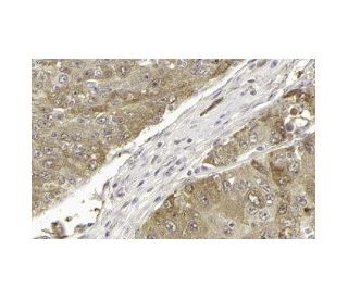
: sc-5295. Colorazione con immunoperossidasi di un tumore del fegato umano fissato in formalina e incluso in paraffina, che mostra la colorazione citoplasmatica delle cellule tumorali. Gentilmente fornito dal programma svedese Human Protein Atlas (HPA).")
: sc-6866. Analisi Western blot dell'espressione di ICAD nel lisato di cellule intere HeLa.")
Anticorpo CAD (H-1): sc-5295
- CAD Antibody H-1 è un monoclonale di topo IgG2b κ, citato in 2 pubblicazioni, fornito in 200 µg/ml
- generato contro gli amminoacidi 1-338 di CAD (caspase-activated deoxyribonuclease) di origine human
- raccomandato per il rilevamento di CAD di origine human in WB, IP, IF, IHC(P) e ELISA
- disponibile coniugato ad agarosio per IP
- m-IgG Fc BP-HRP, 2b BP-HRP">m-IgG2b BP-HRP e m-IgGκ BP-HRP sono i reagenti secondari preferiti per l'anticorpo CAD (H-1) per applicazioni WB e IHC(P). Questi reagenti sono ora offerti in bundle con l'anticorpo CAD (H-1)(vedere le informazioni per l'ordine sotto).

LINK RAPIDI
VEDI ANCHE...
La famiglia delle cisteine proteasi Ced/ICE o caspasi svolge un ruolo fondamentale nel mediare l'apoptosi attraverso la proteolisi di bersagli specifici. Tra i bersagli vi sono la poli(ADP-ribosio) polimerasi (PARP), la gelsolina, l'ICAD (inibitore della CAD, designato anche DFF-45) e le lamine nucleari. La CAD (desossiribonucleasi attivata dalla caspasi), designata anche CPAN (nucleasi attivata dalla caspasi) e DFF40, è una DNasi responsabile della degradazione del DNA durante l'apoptosi. La caspasi-3 agisce per dissociare la CAD dall'ICAD, consentendo alla CAD di entrare nel nucleo e degradare il DNA.
Alexa Fluor® è un marchio registrato di Molecular Probes Inc., OR., USA
LI-COR® e Odyssey® sono marchi registrati di LI-COR Biosciences
Anticorpo CAD (H-1) Riferimenti:
- Rilevamento della scissione del DNA nelle cellule apoptotiche. | Kaufmann, SH., et al. 2000. Methods Enzymol. 322: 3-15. PMID: 10914000
- Possibile coinvolgimento della ciclofilina B e della deossiribonucleasi attivata dalla caspasi nell'induzione della degradazione del DNA cromosomico nei timociti stimolati dal TCR. | Nagata, T., et al. 2000. J Immunol. 165: 4281-9. PMID: 11035062
- La caspasi-3 e la deossiribonucleasi attivata dalla caspasi sono associate all'apoptosi delle cellule germinali testicolari derivante dalla riduzione del testosterone intratesticolare. | Kim, JM., et al. 2001. Endocrinology. 142: 3809-16. PMID: 11517157
- Induzione dell'attività della deossiribonucleasi attivata dalla caspasi dopo ischemia cerebrale focale e riperfusione. | Luo, Y., et al. 2002. J Cereb Blood Flow Metab. 22: 15-20. PMID: 11807389
- Apoptosi sopravvissuta. | Vaughan, AT., et al. 2002. Apoptosis. 7: 173-7. PMID: 11865202
- Attivazione dell'endonucleasi e frammentazione del DNA cromosomico durante l'apoptosi nelle cellule leucemiche. | Yoshida, A., et al. 2006. Int J Hematol. 84: 31-7. PMID: 16867899
- La frammentazione apoptotica del DNA può essere un'attività cooperativa tra la deossiribonucleasi attivata dalla caspasi e la DNAS1L3 regolata dalla poli(ADP-ribosio) polimerasi, un'endonucleasi localizzata nel reticolo endoplasmatico che si trasla nel nucleo durante l'apoptosi. | Errami, Y., et al. 2013. J Biol Chem. 288: 3460-8. PMID: 23229555
- Il collasso della cromatina durante la morte cellulare apoptotica dipendente dalla caspasi richiede il fattore di frammentazione del DNA, rotture del DNA a singolo filamento 3'-OH mediate dalla subunità di 40 kDa/deossiribonucleasi attivata dalla caspasi. | Iglesias-Guimarais, V., et al. 2013. J Biol Chem. 288: 9200-15. PMID: 23430749
- Le terapie mirate con piccole molecole inducono una dipendenza dalla riparazione della rottura del doppio filamento di DNA nelle cellule tumorali residue. | Ali, M., et al. 2022. Sci Transl Med. 14: eabc7480. PMID: 35353542
- Una DNasi attivata dalla caspasi che degrada il DNA durante l'apoptosi e il suo inibitore ICAD. | Enari, M., et al. 1998. Nature. 391: 43-50. PMID: 9422506
Informazioni ordini
| Nome del prodotto | Codice del prodotto | UNITÀ | Prezzo | Quantità | Preferiti | |
CAD Anticorpo (H-1) | sc-5295 | 200 µg/ml | $322.00 | |||
CAD (H-1): m-IgG Fc BP-HRP Bundle | sc-536548 | 200 µg Ab; 10 µg BP | $361.00 | |||
CAD (H-1): m-IgGκ BP-HRP Bundle | sc-533699 | 200 µg Ab; 40 µg BP | $361.00 | |||
CAD (H-1): m-IgG2b BP-HRP Bundle | sc-549809 | 200 µg Ab; 10 µg BP | $361.00 | |||
CAD Anticorpo (H-1) AC | sc-5295 AC | 500 µg/ml, 25% agarose | $424.00 |