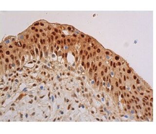
Date published: 2025-12-25

1-800-457-3801

SCBT Portrait Logo
Seach Input

p-Akt1/2/3 Antibody (B-12): sc-377556

4.2(10)
Write a reviewAsk a question

Datasheets
  • p-Akt1/2/3 Antibody (B-12) is a mouse monoclonal IgM κ, cited in 38 publications, provided at 200 µg/ml
  • specific for an epitope mapping between amino acids Thr 450 Thr 450 of Akt1 of human origin
  • recommended for detection of Thr 450 phosphorylated Akt1 and Thr 451 correspondingly phosphorylated Akt2 and Thr 447 correspondingly phosphorylated Akt3 of mouse, rat and human origin by WB, IP, IF, IHC(P) and ELISA; also reactive with additional species, including and equine, bovine and porcine
  • At present, we have not yet completed the identification of the preferred secondary detection reagent(s) for p-Akt1/2/3 Antibody (B-12). This work is in progress.

QUICK LINKS

SEE ALSO...

p-Akt1/2/3 Antibody (B-12) is a mouse monoclonal IgM antibody that detects p-Akt1/2/3 in mouse, rat, and human samples through applications such as western blotting (WB), immunoprecipitation (IP), immunofluorescence (IF), immunohistochemistry, and enzyme-linked immunosorbent assay (ELISA). p-Akt1/2/3 monoclonal antibody (B-12) is available in a non-conjugated form, allowing for versatile experimental use. The Akt family of serine/threonine kinases, which includes Akt1, Akt2, and Akt3, plays a crucial role in various cellular processes, including metabolism, cell survival, and growth. Notably, Akt proteins are activated by growth factors such as insulin and insulin-like growth factor 1 (IGF-1), leading to their phosphorylation at specific threonine and serine residues. This phosphorylation enables Akt to transduce signals from the phosphatidylinositol 3-kinase (PI3K) pathway, which regulates cellular responses to growth signals. The interaction of Akt with other proteins, such as phosphoinositide-dependent kinase 1 (PDK1), is critical for activation and subsequent signaling cascade. Akt3, expressed in tissues like adipocytes and skeletal muscle, is involved in processes such as glucose uptake and glycogen synthesis, highlighting importance in metabolic regulation. p-Akt1/2/3 (B-12) antibody specifically recognizes phosphorylated forms of Akt1, Akt2, and Akt3, making this antibody an invaluable tool for researchers studying signaling pathways that govern cell growth and metabolism.

For Research Use Only. Not Intended for Diagnostic or Therapeutic Use.

Ordering Information

Product NameCatalog #UNITPriceQtyFAVORITES

p-Akt1/2/3 Antibody (B-12)

sc-377556
200 µg/ml
$316.00

p-Akt1/2/3 (B-12) Neutralizing Peptide

sc-377556 P
100 µg/0.5 ml
$68.00

I am using B-12 antibody (pAKT), is it igM second antibody with HRP to be used for detection? or will it work with igGk second antibody?

Asked by: Chai 123
Thank you for your question. p-Akt1/2/3 Antibody (B-12) is a mouse monoclonal IgM and we do recommend using an isotype specific secondary anti-mouse IgM for detection. Alternatively, you could also use our mouse IgG kappa binding protein (m-IgGκ BP) conjugated to Horseradish Peroxidase (HRP), m-IgGκ BP-HRP: sc-516102. This secondary antibody binds specifically to the kappa light chain of mouse IgG, and is also reactive with the kappa light chain of mouse IgM. A link to the product information is provided below for your reference. https://www.scbt.com/scbt/product/m-igg-kappa-bp-hrp
Answered by: Technical Support
Date published: 2017-06-29

What application is the blocking peptide sc-377556 P appropriate for?

Asked by: DefinitelyNotMatt
Thank you for your question. The blocking peptide is intended for use as a negative control, by pre-adsorbing the mouse monoclonal antibody against the antigen. For full protocol details, please contact our Technical Services Department or view our online protocol here: https://www.scbt.com/scbt/resources/protocols/peptide-neutralization
Answered by: Technical Support
Date published: 2017-02-24
  • y_2025, m_12, d_20, h_7CST
  • bvseo_bulk, prod_bvqa, vn_bulk_3.0.42
  • cp_1, bvpage1
  • co_hasquestionsanswers, tq_2
  • loc_en_US, sid_377556, prod, sort_[SortEntry(order=LAST_APPROVED_ANSWER_SUBMISSION_TIME, direction=DESCENDING)]
  • clientName_scbt
  • bvseo_sdk, java_sdk, bvseo-4.0.0
  • CLOUD, getContent, 109ms
  • QUESTIONS, PRODUCT
Rated 4 out of 5 by from Showed specific bandsGood antibody showed specific band with 1:500 dilution.
Date published: 2017-10-20
Rated 4 out of 5 by from Not very sureSample was mixed with 5X Reducing Loading Buffer and Denature the proteins by heating the sample to 95°C. Load the sample onto 3% Stacking Gel and 10% Separation Gel and run the gel under standard conditions. Transfer the proteins to a PVDF membrane using semi-dry or wet transfer methods. Block the membrane with 3% w/v skim milk powder in TBST. Incubate for 2 hr with agitation. Dilute the primary antibody in WB Anti-body Diluent (1:200) to the recommended dilution. Incubate overnight at 4°C, and rinse the blot in TBST four times for 5 min each at room temperature. Dilute the HRP labeled goat anti-mouse secondary antibody at the Second Ab dilute (1/5000). Incubate 1hr at room temperature, and rinse the blot in four times for 5 min each at room temperature. Perform ECL Plus detection. The result shows below: Not bands, hoverever, were showed in the membrane. That may because our lab used chicken sample, the result testified its antibody also cannot apply in chicken sample.
Date published: 2017-10-18
Rated 4 out of 5 by from Good antibodyWorked well with western blotting at 1:500 dilution.
Date published: 2017-09-27
Rated 4 out of 5 by from Gute Ergebnisse in der IHC(P) auf Herz/RatteMit Standardprotokoll ohne AG Demaskierung ON/4°C, mouse IgGkappa BP. Gute Ergebnisse in der IHC(P) auf Herz/Ratte.
Date published: 2017-08-15
Rated 5 out of 5 by from great antibodyThe result is good , although there is background .the band is clear.
Date published: 2017-07-24
Rated 3 out of 5 by from Nice productThere were some slight background but nice product.
Date published: 2017-05-08
Rated 5 out of 5 by from Worked Great!I was given a sample of this and it has worked great!
Date published: 2017-03-27
Rated 3 out of 5 by from Suitable antibody for Western blottingThis monoclonal antibody was independently tested by Kinexus Bioinformatics Corporation and is recommended for Western blotting. There was medium detection of ~ 60 kDa Akt in overnight serum-starved human A431 cells treated for 5 minutes with EGF on Western blots, and no significant cross-reactivities on these blots with any off-target proteins, except for weak-medium ~80, 135 and 150 kDa proteins.
Date published: 2017-03-14
  • y_2025, m_12, d_20, h_7
  • bvseo_bulk, prod_bvrr, vn_bulk_3.0.42
  • cp_1, bvpage1
  • co_hasreviews, tv_0, tr_10
  • loc_en_US, sid_377556, prod, sort_[SortEntry(order=SUBMISSION_TIME, direction=DESCENDING)]
  • clientName_scbt
  • bvseo_sdk, java_sdk, bvseo-4.0.0
  • CLOUD, getReviews, 15ms
  • REVIEWS, PRODUCT
p-Akt1/2/3 Antibody (B-12) is rated 4.2 out of 5 by 10.
  • y_2025, m_12, d_20, h_7
  • bvseo_bulk, prod_bvrr, vn_bulk_3.0.42
  • cp_1, bvpage1
  • co_hasreviews, tv_0, tr_10
  • loc_en_US, sid_377556, prod, sort_[SortEntry(order=SUBMISSION_TIME, direction=DESCENDING)]
  • clientName_scbt
  • bvseo_sdk, java_sdk, bvseo-4.0.0
  • CLOUD, getAggregateRating, 106ms
  • REVIEWS, PRODUCT