")
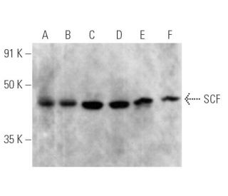
SCF 항체(G-3): sc-13126. MG-63(A), NCI-H1299(B), NIH/3T3(C), Neuro-2A(D), C6(E) 및 PC-12(F) 전체 세포 용해물에서 SCF 발현의 웨스턴 블롯 분석.
SCF 항체 (G-3): sc-13126
- SCF 항체 G-3 는 마우스 monoclonal IgG2b κ SCF 항체, 53간행물에 인용, 이며 200 µg/ml으로 제공합니다.
- human origin의 stem cell factor (SCF)에서 내부에 위치한 26-214 아미노산을 항원으로 사용하였습니다.
- SCF 항체 (G-3)는 WB, IP, IF, IHC(P) and ELISA으로 mouse, rat and human유래의 SCF 를 감지하는 데에 추천한다.
- 항-SCF 항체(G-3)는 IP용 agarose와 결합되어 이용 가능하며, WB, IHC(P), ELISA용 HRP와 결합되어 이용 가능하며, IF, IHC(P), FCM용 phycoerythrin 또는 FITC와 결합되어 이용 가능합니다.
- WB (RGB), IF, IHC(P) 와FCM, RGB fluorescent imaging systems, such as iBright™ FL1000, FluorChem™, Typhoon, Azure and other comparable systems에 사용가능한 Alexa Fluor® 488, Alexa Fluor® 546, Alexa Fluor® 594 or Alexa Fluor® 647결합제품도 있습니다.
- WB (NIR), IF와FCM,Near-Infrared (NIR) detection systems, such as LI-COR®Odyssey®, iBright™ FL1000, FluorChem™, Typhoon, Azure and other comparable systems에 사용가능한 Alexa Fluor® 680 or Alexa Fluor® 790 결합제품도 있습니다.
- 2b BP-HRP">m-IgG2b BP-HRP 및 m-IgGκ BP-HRP는 SCF 항체 (G-3) WB 및 IHC(P) 애플리케이션용입니다. 이 시약은 이제 SCF 항체 (G-3)와 함께 번들로 제공됩니다(아래 주문 정보 참조).

빠른 링크
보조 제품
설명
Gene 정보
프로틴 시퀀스
데이터 시트 및 프로토콜
연구 정보
더보기
SCF 항체(G-3)는 마우스, 랫트, 인간에서 유래된 SCF 단백질을 WB, IP, IF, IHCP, ELISA를 통해 검출하는 마우스 단일 클론 IgG2b 카파 경쇄 항체입니다. SCF 항체(G-3)는 비접합 형태와 아가로스, HRP, PE, FITC, 다중 알렉사 플루오르® 접합체를 포함한 다양한 접합 형태로 제공됩니다. SCF는 여러 세포 과정에 필수적인 막 관통 티로신 키나아제 수용체 원발암 유전자 c-Kit의 리간드로서 중요한 역할을 합니다. SCF는 인간과 쥐의 체내에서 각각 248개와 220개의 아미노산으로 구성되어 있으며, 두 가지의 다른 형태로 존재하는 다형성 사이토카인입니다. 더 큰 형태의 SCF는 주로 섬유세포, 뇌, 흉선에서 발현되는 반면, 더 작은 변이체는 비장, 고환, 태반, 소뇌와 같은 조직에서 발견됩니다. SCF는 성숙 및 미성숙 비만세포의 증식과 성숙을 촉진하기 때문에 생식세포, 조혈모세포, 멜라닌세포 전구세포의 발달에 필수적입니다. SCF와 c-Kit의 상호작용은 조혈과 색소 침착을 포함한 다양한 생물학적 과정에 중요하기 때문에, SCF 단일클론항체(G-3)는 발달 생물학 및 암 연구에 유용한 도구입니다.
연구용으로만 사용가능합니다. 진단이나 치료용으로 사용불가합니다.
Alexa Fluor®는 미국 오리건주 Molecular Probes Inc.의 상표입니다.
LI-COR®와 Odyssey®는 LI-COR Biosciences의 등록 상표입니다.
SCF 항체 (G-3) 참고문헌:
- 두 가지 세포 관련 형태의 키트 리간드에 대한 차별적 발현 및 처리: KL-1 및 KL-2. | Huang, EJ., et al. 1992. Mol Biol Cell. 3: 349-62. PMID: 1378327
- 강철 유전자 산물인 줄기세포 인자의 인간 동종 단백질의 막 결합 및 분비 형태를 선택적으로 발현하는 쥐 기질 세포에 의한 장기 골수 배양에서 인간 조혈 지원. | Toksoz, D., et al. 1992. Proc Natl Acad Sci U S A. 89: 7350-4. PMID: 1380155
- 막 고정 성장 인자의 분열에는 공통 메커니즘을 통해 조절되는 뚜렷한 프로테아제 활동이 수반됩니다. | Pandiella, A., et al. 1992. J Biol Chem. 267: 24028-33. PMID: 1385433
- 비만 세포 성장 인자는 마우스 염색체 10번의 강철 유전자좌 근처에 위치하며 다수의 강철 대립 유전자에서 결실되어 있습니다. | Copeland, NG., et al. 1990. Cell. 63: 175-83. PMID: 1698554
- 쥐와 인간 줄기세포 인자 DNA의 기본 구조 및 기능적 발현. | Martin, FH., et al. 1990. Cell. 63: 203-11. PMID: 2208279
- 태반 틈새에서 줄기세포 인자의 역할. | Khodadi, E., et al. 2016. Cell Tissue Res. 366: 523-531. PMID: 27234501
- 적혈구 생성 인자, 줄기세포 인자, 그리고 암세포 이동. | Vazquez-Mellado, MJ., et al. 2017. Vitam Horm. 105: 273-296. PMID: 28629522
- 골수 지방세포는 SCF를 분비하여 줄기세포의 재생과 조혈을 촉진합니다. | Zhou, BO., et al. 2017. Nat Cell Biol. 19: 891-903. PMID: 28714970
- 줄기세포 인자: 골수 지방세포와 조혈세포 사이의 다리. | Li, Z. and MacDougald, OA. 2019. Haematologica. 104: 1689-1691. PMID: 31473604
- 가용성 줄기세포 인자의 억제는 장 점막의 회복을 촉진합니다. | Garcia-Hernandez, V., et al. 2023. Inflamm Bowel Dis. 29: 1133-1144. PMID: 36688460
- 줄기세포 인자와 cKIT는 저산소 상태에서 내피세포의 당분해 작용을 조절합니다. | Jeong, H., et al. 2024. Cardiovasc Res. 120: 745-755. PMID: 38507654
- 단핵구는 SCF가 있는 상태에서 배양할 때 비만세포를 만들지 않습니다. 순환하는 비만세포 전구세포를 c-kit+, CD34+, Ly-, CD14-, CD17-, 콜로니 형성 세포로 특성화합니다. | Agis, H., et al. 1993. J Immunol. 151: 4221-7. PMID: 7691941
주문정보
| 제품명 | 카탈로그 번호 | 단위 | 가격 | 수량 | 관심품목 | |
SCF 항체 (G-3) | sc-13126 | 200 µg/ml | $322.00 | |||
SCF (G-3): m-IgGκ BP-HRP 번들 | sc-520598 | 200 µg Ab, 40 µg BP | $361.00 | |||
SCF (G-3): m-IgG2b BP-HRP 번들 | sc-548841 | 200 µg Ab; 10 µg BP | $361.00 | |||
SCF 항체 (G-3) AC | sc-13126 AC | 500 µg/ml, 25% agarose | $424.00 | |||
SCF 항체 (G-3) HRP | sc-13126 HRP | 200 µg/ml | $322.00 | |||
SCF 항체 (G-3) FITC | sc-13126 FITC | 200 µg/ml | $336.00 | |||
SCF 항체 (G-3) PE | sc-13126 PE | 200 µg/ml | $349.00 | |||
SCF 항체 (G-3) Alexa Fluor® 488 | sc-13126 AF488 | 200 µg/ml | $364.00 | |||
SCF 항체 (G-3) Alexa Fluor® 546 | sc-13126 AF546 | 200 µg/ml | $364.00 | |||
SCF 항체 (G-3) Alexa Fluor® 594 | sc-13126 AF594 | 200 µg/ml | $364.00 | |||
SCF 항체 (G-3) Alexa Fluor® 647 | sc-13126 AF647 | 200 µg/ml | $364.00 | |||
SCF 항체 (G-3) Alexa Fluor® 680 | sc-13126 AF680 | 200 µg/ml | $364.00 | |||
SCF 항체 (G-3) Alexa Fluor® 790 | sc-13126 AF790 | 200 µg/ml | $364.00 |