")
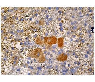
: sc-365943. Colorazione con immunoperossidasi di tessuto di ghiandola surrenale umana fissato in formalina e incluso in paraffina che mostra la colorazione citoplasmatica di un sottogruppo di cellule ghiandolari.")
: sc-365943. Analisi Western blot dell'espressione di Ret in lisati di cellule intere 293T non trasfettate: sc-117752 (A) e trasfettate con Ret umano: sc-158925 (B).")
Anticorpo Ret (C-3): sc-365943
- Ret Antibody C-3 è un monoclonale di topo IgG1 κ Ret antibody, citato in 5 pubblicazioni, fornito in 200 µg/ml
- generato contro gli amminoacidi 31-330 mappati vicino al N-terminus di a region conserved between Ret isoforms A and C di origine human
- Ret Antibody (C-3) é raccomandato per il rilevamento di Ret isoforms A and C di origine human in WB, IP, IF, IHC(P) e ELISA
- Ret Antibody (C-3) é disponibile coniugato con agarose per IP; HRP per WB, IHC(P) ed ELISA; sia con phycoerythrin o FITC per IF, IHC(P) e FCM
- disponibile anche coniugato con Alexa Fluor® 488, Alexa Fluor® 546; Alexa Fluor® 594 o Alexa Fluor® 647 per IF, IHC (P) e FCM
- disponibile anche coniugato con Alexa Fluor® 680 o Alexa Fluor® 790 per WB (NIR), IF e FCM
- m-IgG Fc BP-HRP, 1 BP-HRP">m-IgG1 BP-HRP e m-IgGκ BP-HRP sono i reagenti secondari preferiti per l'anticorpo Ret (C-3) per applicazioni WB e IHC(P). Questi reagenti sono ora offerti in bundle con l'anticorpo Ret (C-3)(vedere le informazioni per l'ordine sotto).

LINK RAPIDI
VEDI ANCHE...
L'anticorpo Ret (C-3) è un anticorpo monoclonale IgG1 κ di topo che rileva la proteina Ret di origine umana mediante WB, IP, IF, IHC(P) ed ELISA. L'anticorpo Ret (C-3) è disponibile sia nella forma anticorpo anti-Ret non coniugato, sia in molteplici forme coniugate di anticorpo anti-Ret, tra cui agarosio, HRP, PE, FITC e molteplici coniugati Alexa Fluor®. Il proto-oncogene Ret è strutturalmente correlato alla crescente famiglia di recettori transmembrana tirosin-chinasi ed è coinvolto nella segnalazione del GDNF. Mediante splicing alternativo, vengono generate due isoforme del proto-oncogene Ret. Le isoforme differiscono tra loro per avere 9 o 51 aminoacidi carbossilici terminali. I prodotti del gene Ret comprendono due proteine glicosilate e, nelle cellule trattate con tunicamicina, una proteina non glicosilata coerente con il peso molecolare Ret previsto in base all'analisi della sequenza. Nei carcinomi papillari della tiroide sono stati identificati riarrangiamenti tumorali specifici del proto-oncogene Ret che portano alla formazione di diverse proteine di fusione trasformanti che condividono il dominio tirosin-chinasico di Ret. A differenza del proto-oncogene Ret, le forme riarrangiate sono costitutivamente fosforilate sulla tirosina e vengono traslocate dalla membrana al citoplasma.
Alexa Fluor® è un marchio registrato di Molecular Probes Inc., OR., USA
LI-COR® e Odyssey® sono marchi registrati di LI-COR Biosciences
Anticorpo Ret (C-3) Riferimenti:
- Identificazione del prodotto di due forme oncogene riarrangiate del proto-oncogene RET nei carcinomi papillari della tiroide. | Lanzi, C., et al. 1992. Oncogene. 7: 2189-94. PMID: 1437145
- Identificazione dei prodotti del proto-oncogene ret nelle cellule di neuroblastoma e di leucemia. | Takahashi, M., et al. 1991. Oncogene. 6: 297-301. PMID: 2000222
- Caratterizzazione degli mRNA del proto-oncogene ret che codificano due isoforme del prodotto proteico in una linea cellulare di neuroblastoma umano. | Tahira, T., et al. 1990. Oncogene. 5: 97-102. PMID: 2181380
- Il PTC è una nuova forma riarrangiata del proto-oncogene ret ed è frequentemente rilevato in vivo nei carcinomi papillari tiroidei umani. | Grieco, M., et al. 1990. Cell. 60: 557-63. PMID: 2406025
- Crosstalk di parkina e Ret nei neuroni dopaminergici. | Kramer, ER. 2015. Oncotarget. 6: 15704-5. PMID: 26202959
- Clonazione ed espressione del proto-oncogene ret che codifica una tirosina chinasi con due potenziali domini transmembrana. | Takahashi, M., et al. 1988. Oncogene. 3: 571-8. PMID: 3078962
- Terapie mirate per il cancro da fusione RET: Dilemmi e progressi. | Ding, S., et al. 2020. Biomed Pharmacother. 132: 110901. PMID: 33125973
- Caratteri distintivi di RET e alterazioni genomiche coincidenti nei tumori RET-aberranti. | Adashek, JJ., et al. 2021. Mol Cancer Ther. 20: 1769-1776. PMID: 34493590
- L'evoluzione della resistenza agli inibitori di RET nei tumori polmonari e tiroidei RET-driven. | Rosen, EY., et al. 2022. Nat Commun. 13: 1450. PMID: 35304457
- RET è un regolatore sessuale della tumorigenesi intestinale. | Koester, ST., et al. 2023. Front Gastroenterol (Lausanne). 2: PMID: 39148929
- Recettore funzionale per GDNF codificato dal proto-oncogene c-ret. | Trupp, M., et al. 1996. Nature. 381: 785-9. PMID: 8657281
- Mutazioni RET nelle malattie umane. | Pasini, B., et al. 1996. Trends Genet. 12: 138-44. PMID: 8901418
Informazioni ordini
| Nome del prodotto | Codice del prodotto | UNITÀ | Prezzo | Quantità | Preferiti | |
Ret Anticorpo (C-3) | sc-365943 | 200 µg/ml | $322.00 | |||
Ret (C-3): m-IgG Fc BP-HRP Bundle | sc-529522 | 200 µg Ab; 10 µg BP | $361.00 | |||
Ret (C-3): m-IgGκ BP-HRP Bundle | sc-522436 | 200 µg Ab, 40 µg BP | $361.00 | |||
Ret (C-3): m-IgG1 BP-HRP Bundle | sc-543559 | 200 µg Ab; 20 µg BP | $361.00 | |||
Ret Anticorpo (C-3) AC | sc-365943 AC | 500 µg/ml, 25% agarose | $424.00 | |||
Ret Anticorpo (C-3) HRP | sc-365943 HRP | 200 µg/ml | $322.00 | |||
Ret Anticorpo (C-3) FITC | sc-365943 FITC | 200 µg/ml | $336.00 | |||
Ret Anticorpo (C-3) PE | sc-365943 PE | 200 µg/ml | $349.00 | |||
Ret Anticorpo (C-3) Alexa Fluor® 488 | sc-365943 AF488 | 200 µg/ml | $364.00 | |||
Ret Anticorpo (C-3) Alexa Fluor® 546 | sc-365943 AF546 | 200 µg/ml | $364.00 | |||
Ret Anticorpo (C-3) Alexa Fluor® 594 | sc-365943 AF594 | 200 µg/ml | $364.00 | |||
Ret Anticorpo (C-3) Alexa Fluor® 647 | sc-365943 AF647 | 200 µg/ml | $364.00 | |||
Ret Anticorpo (C-3) Alexa Fluor® 680 | sc-365943 AF680 | 200 µg/ml | $364.00 | |||
Ret Anticorpo (C-3) Alexa Fluor® 790 | sc-365943 AF790 | 200 µg/ml | $364.00 |