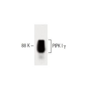
Date published: 2026-6-6

1-800-457-3801

SCBT Portrait Logo
Seach Input

PIPK I γ Antibody (12): sc-136033

4.0(1)
Write a reviewAsk a question

Datasheets
  • PIPK I γ Antibody (12) is a mouse monoclonal IgG1 provided at 50 µg/0.5 ml
  • raised against amino acids 479-580 of PIPK I γ of mouse origin
  • recommended for detection of PIPK I γ of mouse and rat origin by WB, IP and IF
  • At present, we have not yet completed the identification of the preferred secondary detection reagent(s) for PIPK I γ Antibody (12). This work is in progress.
    Gene Editing Promo Banner

    QUICK LINKS

    SEE ALSO...

    PIPK I γ Antibody (12) is a mouse monoclonal IgG1 antibody that detects PIPK I γ in mouse and rat samples through applications such as western blotting (WB), immunoprecipitation (IP), and immunofluorescence (IF). PIPK I γ plays a crucial role in cellular signaling by synthesizing phosphatidylinositol-4,5-bisphosphate, a key phospholipid that regulates various cellular processes, including cell proliferation, survival, and cytoskeletal organization. PIPK I γ is involved in modulating membrane trafficking and organizing the cytoskeleton, which are essential for maintaining cellular integrity and facilitating communication between cells. PIPK I γ is expressed in several tissues, including the lung, brain, and kidneys, and exists in two variants produced by alternative splicing, highlighting functional diversity and significance in different biological contexts. The PIPK family, which includes type I, type II, and type III kinases, is characterized by distinct substrate specificity and subcellular targeting, with the activation loop playing a pivotal role in determining these properties. PIPK I γ, along with its family members, underscores the complexity of phosphoinositide signaling pathways and their implications in health and disease, making anti-PIPK I γ antibody (12) an invaluable tool for researchers studying these critical processes.

    For Research Use Only. Not Intended for Diagnostic or Therapeutic Use.

    Alexa Fluor® is a trademark of Molecular Probes Inc., OR., USA

    LI-COR® and Odyssey® are registered trademarks of LI-COR Biosciences

    PIPK I γ Antibody (12) References:

    1. PIPKIγ regulates β-catenin transcriptional activity downstream of growth factor receptor signaling.  |  Schramp, M., et al. 2011. Cancer Res. 71: 1282-91. PMID: 21303971
    2. PIPKIγ regulates focal adhesion dynamics and colon cancer cell invasion.  |  Wu, Z., et al. 2011. PLoS One. 6: e24775. PMID: 21931851
    3. PIPKIγ targets to the centrosome and restrains centriole duplication.  |  Xu, Q., et al. 2014. J Cell Sci. 127: 1293-305. PMID: 24434581
    4. Isoform 5 of PIPKIγ regulates the endosomal trafficking and degradation of E-cadherin.  |  Schill, NJ., et al. 2014. J Cell Sci. 127: 2189-203. PMID: 24610942
    5. Cross-Talk between Shp1 and PIPKIγ Controls Leukocyte Recruitment.  |  Stadtmann, A., et al. 2015. J Immunol. 195: 1152-61. PMID: 26101325
    6. Phosphatidylinositol phosphate kinase PIPKIγ and phosphatase INPP5E coordinate initiation of ciliogenesis.  |  Xu, Q., et al. 2016. Nat Commun. 7: 10777. PMID: 26916822
    7. FAK, talin and PIPKIγ regulate endocytosed integrin activation to polarize focal adhesion assembly.  |  Nader, GP., et al. 2016. Nat Cell Biol. 18: 491-503. PMID: 27043085
    8. PIPKIγ and talin couple phosphoinositide and adhesion signaling to control the epithelial to mesenchymal transition.  |  Thapa, N., et al. 2017. Oncogene. 36: 899-911. PMID: 27452517
    9. PIPKIγ Regulates CCL2 Expression in Colorectal Cancer by Activating AKT-STAT3 Signaling.  |  Xue, J., et al. 2019. J Immunol Res. 2019: 3690561. PMID: 31781676
    10. Ciliopathy protein HYLS1 coordinates the biogenesis and signaling of primary cilia by activating the ciliary lipid kinase PIPKIγ.  |  Chen, C., et al. 2021. Sci Adv. 7: PMID: 34162535

    Ordering Information

    Product NameCatalog #UNITPriceQtyFAVORITES

    PIPK I γ Antibody (12)

    sc-136033
    50 µg/0.5 ml
    $322.00

    I am using PIPK I γ (12): sc-136033 for immunofluorescence, what dilution of this antibody should I use?

    Asked by: Germaine
    Thank you for your question. We recommend a starting dilution of 1:50. However, optimal antibody concentration should be determined by titration. Please find our full immunofluorescence protocol here: https://www.scbt.com/scbt/resources/protocols/immunofluorescence-cell-staining
    Answered by: Technical Support
    Date published: 2017-03-24
    • y_2026, m_6, d_4, h_10CST
    • bvseo_bulk, prod_bvqa, vn_bulk_3.0.46
    • cp_1, bvpage1
    • co_hasquestionsanswers, tq_1
    • loc_en_US, sid_136033, prod, sort_[SortEntry(order=LAST_APPROVED_ANSWER_SUBMISSION_TIME, direction=DESCENDING)]
    • clientName_scbt
    • bvseo_sdk, java_sdk, bvseo-4.0.0
    • CLOUD, getContent, 81ms
    • QUESTIONS, PRODUCT
    Rated 4 out of 5 by from Very strong band of interest observed inVery strong band of interest observed in WB in rat cerebrum tissue extract. -SCBT QC
    Date published: 2013-02-15
    • y_2026, m_6, d_4, h_10
    • bvseo_bulk, prod_bvrr, vn_bulk_3.0.46
    • cp_1, bvpage1
    • co_hasreviews, tv_0, tr_1
    • loc_en_US, sid_136033, prod, sort_[SortEntry(order=SUBMISSION_TIME, direction=DESCENDING)]
    • clientName_scbt
    • bvseo_sdk, java_sdk, bvseo-4.0.0
    • CLOUD, getReviews, 7ms
    • REVIEWS, PRODUCT
    PIPK I γ Antibody (12) is rated 4.0 out of 5 by 1.
    • y_2026, m_6, d_4, h_10
    • bvseo_bulk, prod_bvrr, vn_bulk_3.0.46
    • cp_1, bvpage1
    • co_hasreviews, tv_0, tr_1
    • loc_en_US, sid_136033, prod, sort_[SortEntry(order=SUBMISSION_TIME, direction=DESCENDING)]
    • clientName_scbt
    • bvseo_sdk, java_sdk, bvseo-4.0.0
    • CLOUD, getAggregateRating, 78ms
    • REVIEWS, PRODUCT