
Date published: 2026-10-28

1-800-457-3801

SCBT Portrait Logo
Seach Input

p-p53 항체 (FP3.2): sc-51690

4.2(14)
리뷰 쓰기질문하기
데이터 시트
  • p-p53 항체(FP3.2) 는 마우스 monoclonal IgG1κ, 17간행물에 인용, 이며 100 µg/ml으로 제공합니다.
  • human 기원의 p53의 아미노산 Ser 392에 해당하는 C-terminal 포스포펩티드에 대해 배양
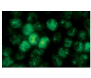
  • WB, IP, IF, IHC(P)와 ELISA으로 mouse, rat와 human origin의 Ser 392 phosphorylated p53을 검출할것을 권장합니다.; dephosphorylated p53 감지에 권장되지 않음
  • m-IgG Fc BP-HRP 및 m-IgG1 BP-HRP 는 p-p53 항체 (FP3.2) for WB and IHC(P) applications. 이 시약은 이제 p-p53 항체 (FP3.2)와 함께 번들로 제공됩니다(아래 주문 정보 참조).

Gene Editing Promo Banner

빠른 링크

더보기

p-p53 항체(FP3.2)는 WB, IP, IF, IHC(P) 및 ELISA에 의해 마우스, 랫 및 인간 기원의 Ser 392 인산화된 p53을 검출하는 IgG1 κ 마우스 단일 클론 p-p53 항체(TP53 항체로도 지정됨)이다. p-p53 항체(FP3.2)는 비결합 항-p53 항체 형태로 사용할 수 있다. p53은 스트레스 신호에 반응하여 성장 정지 및 세포 자멸 관련 유전자를 상향 조절하여 프로그램된 세포 사멸, 세포 분화 및 세포 주기 제어 메커니즘에 영향을 미치는 DNA 결합, 올리고머화 도메인 및 전사 활성화 도메인 포함 종양 억제제이다. p53은 핵에 국한되지만 MDM2 폴리유비퀴틴화 단백질 분해를 위해 활성 p53이 있는 상태에서 상향 조절되는 E3 유비퀴틴 리가아제인 음성 조절기 MDM2에 의해 세포질로 샤페론될 수 있다. p53은 DNA가 없을 때 사량체로 조립될 수 있고, 잠재 및 활성 (DNA 결합) 형태 사이에서 변동하며, 인산화 및 아세틸화를 포함한 번역 후 변형을 통해 차등적으로 활성화된다. p53의 DNA 결합 도메인 (DBD) (아미노산 110-286)의 돌연변이는 시스 요소와 에너지적으로 유리한 연관성을 손상시킬 수 있으며 여러 인간 암과 관련이 있다.

연구용으로만 사용가능합니다. 진단이나 치료용으로 사용불가합니다.

Alexa Fluor®는 미국 오리건주 Molecular Probes Inc.의 상표입니다.

LI-COR®와 Odyssey®는 LI-COR Biosciences의 등록 상표입니다.

p-p53 항체 (FP3.2) 참고문헌:

  1. 쥐에서 화학적 간암 발생 초기 단계의 GST-P, p53 및 p21(ras) 단백질 발현과 항염증 활성에 대한 보쉬니키아 로시카의 효과.  |  Yin, ZZ., et al. 2000. World J Gastroenterol. 6: 812-818. PMID: 11819701
  2. Bcl-2의 상향 조절과 P-p53의 하향 조절은 오리도닌에 의한 세포 사멸에 대한 쥐의 L929 섬유육종 세포의 낮은 민감성을 설명합니다.  |  Huang, J., et al. 2005. Biol Pharm Bull. 28: 2068-74. PMID: 16272691
  3. USP10은 p53의 디유비퀴틴화를 통해 p53의 국소화와 안정성을 조절합니다.  |  Yuan, J., et al. 2010. Cell. 140: 384-96. PMID: 20096447
  4. 디메틸푸마레이트는 MSK1과 RSK1의 활성화를 억제하고 핵 내 p-c-Jun(S63)과 p-p53(S15)의 발현을 유도함으로써 MIF에 의한 각질세포의 증식을 억제합니다.  |  Gesser, B., et al. 2011. Inflamm Res. 60: 643-53. PMID: 21340650
  5. ATM은 DNA 손상에 대한 반응으로 p-p53 및 p-ERK와 삼원 복합체를 형성함으로써 p53과 ERK의 상호 의존적 활성화를 매개합니다.  |  Heo, JI., et al. 2012. Mol Biol Rep. 39: 8007-14. PMID: 22576881
  6. p53과 수모세포종.  |  Ramaswamy, V., et al. 2015. Cold Spring Harb Perspect Med. 6: a026278. PMID: 26684332
  7. 자가포식 작용과 p53.  |  White, E. 2016. Cold Spring Harb Perspect Med. 6: a026120. PMID: 27037419
  8. ATM 키나아제 억제제 KU-55933은 칼파인과 카셉신 D의 억제라는 γH2AX/p-p53/카스파제-3 독립적 메커니즘을 통해 과산화수소로 인한 세포 손상에 대한 신경 보호 기능을 제공합니다.  |  Chwastek, J., et al. 2017. Int J Biochem Cell Biol. 87: 38-53. PMID: 28341201
  9. PKA/p-p53/Bax 신호 경로와 관련된 해리 후 hESC에서 활성화된 미토콘드리아 세포 사멸.  |  Zhang, L., et al. 2018. Exp Cell Res. 369: 226-233. PMID: 29802837
  10. 핵 HKII-P-p53(Ser15) 상호작용은 상피성 난소암에서 화학적 감수성 및 당분해 조절에 대한 예후 바이오마커입니다.  |  Han, CY., et al. 2021. Cancers (Basel). 13: PMID: 34298618

주문정보

제품명카탈로그 번호 단위가격수량관심품목

p-p53 항체 (FP3.2)

sc-51690
100 µg/ml
$322.00

p-p53 (FP3.2): m-IgG Fc BP-HRP 번들

sc-538943
100 µg Ab; 10 µg BP
$361.00

p-p53 (FP3.2): m-IgG1 BP-HRP 번들

sc-541169
100 µg Ab; 20 µg BP
$361.00

What is the recommended starting dilution for Western blot for p-p53 (FP3.2): sc-51690 monoclonal antibody?

Asked by: Randy McDonald
The recommended starting dilution for Western blot is 1:100 with a dilution range of 1:100-1:1000. This information, along with other technical specifications, is available in the recommended protocol: https://www.scbt.com/scbt/resources/protocols/western-immuno-blotting
Answered by: Technical Support
Date published: 2017-04-05
  • y_2026, m_10, d_28, h_5CST
  • bvseo_bulk, prod_bvqa, vn_bulk_3.0.48
  • cp_1, bvpage1
  • co_hasquestionsanswers, tq_1
  • loc_ko_KR, sid_51690, prod, sort_[SortEntry(order=LAST_APPROVED_ANSWER_SUBMISSION_TIME, direction=DESCENDING)]
  • clientName_scbt
  • bvseo_sdk, java_sdk, bvseo-4.0.0
  • CLOUD, getContent, 110ms
  • QUESTIONS, PRODUCT
Rated 4 out of 5 by KuanYH from .Excellent.This antibody works great for western blotting.Excellent.
Date published: 2019-07-29
Rated 5 out of 5 by Fabi from Function good for Western Blot (human samples)I used for the detection of p53 after the induction of DNA damage in Molt4 cells (human cell line). I did not detect in the control samples (without treatment) but after the induction of DNA damage I could detect the presence of phosphorylated p53. I used for western blot (dilution 1/1000).
Date published: 2018-02-19
Rated 5 out of 5 by hluo1 from p-p53 Antibody (FP3.2) very good!strong and clean p-p53 band in human cells treated with DNA damage agents by western blot using a free sample antibody
Date published: 2017-12-08
Rated 4 out of 5 by Just15 from bands at the right size on WBseems to be working for western - samples were human cells in culture
Date published: 2017-11-21
Rated 4 out of 5 by wenzhuzi from for western blotThis antibody did not work well in mESC samples with 1: 500 dilution for western blot.
Date published: 2017-10-10
Rated 5 out of 5 by krystal614 from goodI used this sample antibody a month ago and am so happy that I did western blot and got a good result.
Date published: 2017-09-15
Rated 5 out of 5 by JiHu from Good antibodyThe antibody Works very well for western blot in our rat lungs tissue
Date published: 2017-08-21
Rated 5 out of 5 by its a secret from 好用I get the sample two months ago ,it is very function at WB
Date published: 2017-08-09
  • y_2026, m_10, d_28, h_5
  • bvseo_bulk, prod_bvrr, vn_bulk_3.0.48
  • cp_1, bvpage1
  • co_hasreviews, tv_0, tr_14
  • loc_ko_KR, sid_51690, prod, sort_[SortEntry(order=SUBMISSION_TIME, direction=DESCENDING)]
  • clientName_scbt
  • bvseo_sdk, java_sdk, bvseo-4.0.0
  • CLOUD, getReviews, 9ms
  • REVIEWS, PRODUCT
p-p53 항체 (FP3.2) is rated 4.2 out of 5 by 14.
  • y_2026, m_10, d_28, h_5
  • bvseo_bulk, prod_bvrr, vn_bulk_3.0.48
  • cp_1, bvpage1
  • co_hasreviews, tv_0, tr_14
  • loc_ko_KR, sid_51690, prod, sort_[SortEntry(order=SUBMISSION_TIME, direction=DESCENDING)]
  • clientName_scbt
  • bvseo_sdk, java_sdk, bvseo-4.0.0
  • CLOUD, getAggregateRating, 104ms
  • REVIEWS, PRODUCT