Date published: 2025-9-9

001 800-1338-3838

SCBT Portrait Logo
Seach Input

p-Akt1/2/3 항체 (B-5): sc-271966

4.2(40)
리뷰 쓰기질문하기

데이터 시트
  • p-Akt1/2/3 항체(B-5) 는 마우스 monoclonal IgG2bκ, 114간행물에 인용, 이며 200 µg/ml으로 제공합니다.
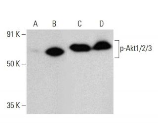
  • p-Akt1/2/3 항체 (B-5)는 WB, IP, IF, IHC(P) and ELISA으로mouse, rat, human and 유래의 Thr 308 phosphorylated Akt1 and correspondingly Thr 309 phosphorylated Akt2 and correspondingly Thr 305 phosphorylated Akt3 를 감지하는 데에 추천한다 ; 이외에 equine and avian등 species와 반응할수 있습니다
  • 항-p-Akt1/2/3 항체(B-5)는 IP용 agarose와 결합되어 이용 가능하며, WB, IHC(P), ELISA용 HRP와 결합되어 이용 가능하며, IF, IHC(P), FCM용 phycoerythrin 또는 FITC와 결합되어 이용 가능합니다.
  • WB (RGB), IF, IHC(P) 와FCM, RGB fluorescent imaging systems, such as iBright™ FL1000, FluorChem™, Typhoon, Azure and other comparable systems에 사용가능한 Alexa Fluor® 488, Alexa Fluor® 546, Alexa Fluor® 594 or Alexa Fluor® 647결합제품도 있습니다.
  • WB (NIR), IF와FCM,Near-Infrared (NIR) detection systems, such as LI-COR®Odyssey®, iBright™ FL1000, FluorChem™, Typhoon, Azure and other comparable systems에 사용가능한 Alexa Fluor® 680 or Alexa Fluor® 790 결합제품도 있습니다.
  • p-Akt1/2/3 (B-5): sc-271966무료10 µg 샘플을 신청하려면 기술지원팀 (또는 로컬 디스트리뷰터)에 연락주세요.
  • m-IgG2b BP-HRP 는p-Akt1/2/3 항체 (B-5) 가 WB and IHC(P) 에 응용될 때 우선으로 사용하는 이차 항체 검사 시약입니다.이 시약은p-Akt1/2/3 항체(B-5) 와 세트로 제공드립니다. (아래의 주문 정보를 참고해 주시길 바랍니다).

빠른 링크

더보기

p-Akt1/2/3 항체(B-5)는 마우스, 쥐 및 인간 유래의 Thr 308 인산화 Akt1 및 이에 상응하는 Thr 309 인산화 Akt2 및 이에 상응하는 Thr 305 인산화 Akt3를 WB, IP, IF, IHC(P) 및 ELISA로 검출하는 IgG2b κ 마우스 모노클로널 p-Akt1/2/3 항체입니다. p-Akt1/2/3 항체(B-5)는 비접합 항-p-Akt1/2/3 항체 형태뿐만 아니라 아가로스, HRP, PE, FITC 및 여러 Alexa Fluor® 접합체를 포함한 여러 접합 형태의 항-p-Akt1/2/3 항체 형태로도 사용할 수 있습니다. 세린/트레오닌 키나아제 Akt 계열에는 단백질 키나아제 A 및 C 계열과 서열 상동성을 나타내며 c-Akt 원시 종양 유전자에 의해 암호화되는 Akt1(PKB 또는 RacPK), Akt2(PKBβ 또는 RacPK-β) 및 Akt 3(PKBγ 또는 thyoma 바이러스 원형 종양 유전자 3) 등 여러 멤버가 포함되어 있습니다. Akt 계열의 모든 구성원은 플렉스트린 상동성 도메인을 가지고 있습니다. Akt1과 Akt2는 PDGF 자극에 의해 활성화됩니다. 이 활성화는 포스파티딜이노시톨 3-키나아제(PI 3-키나아제) 복합체의 서브유닛과 결합하는 PDGFR-β 티로신 잔기 740 및 751에 의존합니다. 인슐린 또는 인슐린 성장 인자-1(IGF-1)에 의한 Akt1의 활성화는 Thr 308과 Ser 473의 인산화를 초래합니다. Akt 단백질은 인슐린/IGF-1 자극 세포에서 업스트림 키나제에 의해 인산화되고 활성화되며, PI 키나제 억제제인 워트만닌에 의해 Akt1 및 Akt2의 활성화가 억제됩니다. 이 데이터를 종합하면 이 단백질이 PI 키나아제의 하류에서 신호를 전달한다는 것을 강력하게 시사합니다. Akt3는 인슐린에 반응하여 세린 잔기에서 인산화됩니다. 그러나 인슐린에 의한 Akt3의 활성화는 PH 도메인의 존재를 필요로 하지 않는 메커니즘을 통해 단백질 키나아제 C의 사전 활성화에 의해 억제됩니다. Akt3는 3T3-L1 섬유아세포, 지방세포 및 골격근에서 발현되며 지방세포 및 근육 분화, 글리코겐 합성, 포도당 흡수, 세포 자멸사 및 세포 증식을 포함한 다양한 생물학적 과정에 관여할 수 있습니다.

연구용으로만 사용가능합니다. 진단이나 치료용으로 사용불가합니다.

Alexa Fluor®는 미국 오리건주 Molecular Probes Inc.의 상표입니다.

LI-COR®와 Odyssey®는 LI-COR Biosciences의 등록 상표입니다.

p-Akt1/2/3 항체 (B-5) 참고문헌:

  1. 세포질 Skp2 발현은 p-Akt1과 관련이 있으며, 인간 유방암의 예후가 좋지 않음을 예측합니다.  |  Liu, J., et al. 2012. PLoS One. 7: e52675. PMID: 23300741
  2. Akt 억제제로서의 4-(피페라진-1-일)-7H-피롤로[2,3-d]피리미딘 유도체의 발견.  |  Liu, Y., et al. 2016. Arch Pharm (Weinheim). 349: 356-62. PMID: 26991997
  3. 슬러그는 p21/p27 단백질을 상향 조절하고, Akt1/p-Akt1 발현을 통한 Wnt/β-카테닌 신호 전달 경로의 활동을 하향 조절함으로써, 인간 자궁 경부암 세포의 증식과 종양 형성을 억제합니다.  |  Cui, N., et al. 2016. Oncotarget. 7: 26152-67. PMID: 27036045
  4. 케르세틴은 p-Akt1, 매트릭스 메탈로프로테이나제(MMP) MMP-2, MMP-9의 발현을 억제함으로써 HCCLM3 세포의 이동과 침입을 억제합니다.  |  Lu, J., et al. 2018. Med Sci Monit. 24: 2583-2589. PMID: 29701200
  5. P-AKT2/SPK1(P-SPK1) 및 P-MEK/P-ERK 세포 신호 전달 경로는 LPS에 의한 대식세포 이동에 관여한다.  |  Lei, Y., et al. 2019. Am J Transl Res. 11: 2725-2741. PMID: 31217849
  6. 다발성 경화증 환자의 말초 T 세포 서브세트에서 Akt1 및 p-Akt1의 발현.  |  Oktelik, FB., et al. 2021. Acta Neurol Belg. 121: 1777-1782. PMID: 33034831
  7. 헥소키나제 2는 자궁경부암 세포에서 Akt1/p-Akt1을 통해 피브로넥틴을 증가시켜 세포 운동성과 원거리 전이를 촉진합니다.  |  Chen, Q., et al. 2021. Cancer Cell Int. 21: 600. PMID: 34758823
  8. 헥소키나제 2는 인간 난소암 세포에서 Akt1/p-Akt1을 활성화하여 세포 운동성과 증식을 촉진합니다.  |  Tian, X., et al. 2022. J Ovarian Res. 15: 92. PMID: 35953860
  9. 호르몬 수용체 양성 및 인간 표피 성장 인자 수용체 2 양성 조기 유방암 환자에서 예후 지표 및 치료 표적으로서의 p-AKT1의 중요성.  |  Kim, JY., et al. 2022. J Breast Cancer. 25: 387-403. PMID: 36314765
  10. IL-21/23 축은 p-Akt1 신호를 통해 RA CD4+ T 세포에서 염증성 사이토카인과 RANKL 발현을 조절합니다.  |  Bhattacharya, G., et al. 2023. Front Immunol. 14: 1235514. PMID: 37809066

주문정보

제품명카탈로그 번호 단위가격수량관심품목

p-Akt1/2/3 항체 (B-5)

sc-271966
200 µg/ml
$316.00

p-Akt1/2/3 (B-5): m-IgG2b BP-HRP 번들

sc-548711
200 µg Ab; 10 µg BP
$354.00

p-Akt1/2/3 항체 (B-5) AC

sc-271966 AC
500 µg/ml, 25% agarose
$416.00

p-Akt1/2/3 항체 (B-5) HRP

sc-271966 HRP
200 µg/ml
$316.00

p-Akt1/2/3 항체 (B-5) FITC

sc-271966 FITC
200 µg/ml
$330.00

p-Akt1/2/3 항체 (B-5) PE

sc-271966 PE
200 µg/ml
$343.00

p-Akt1/2/3 항체 (B-5) Alexa Fluor® 488

sc-271966 AF488
200 µg/ml
$357.00

p-Akt1/2/3 항체 (B-5) Alexa Fluor® 546

sc-271966 AF546
200 µg/ml
$357.00

p-Akt1/2/3 항체 (B-5) Alexa Fluor® 594

sc-271966 AF594
200 µg/ml
$357.00

p-Akt1/2/3 항체 (B-5) Alexa Fluor® 647

sc-271966 AF647
200 µg/ml
$357.00

p-Akt1/2/3 항체 (B-5) Alexa Fluor® 680

sc-271966 AF680
200 µg/ml
$357.00

p-Akt1/2/3 항체 (B-5) Alexa Fluor® 790

sc-271966 AF790
200 µg/ml
$357.00

p-Akt1/2/3 (B-5) 중화펩타이드

sc-271966 P
100 µg/0.5 ml
$68.00

Have you or do you know of western data for use of sc-271966 to detect mouse brain p-Akt1/2/3?

Asked by: Billf
Thank you for your question. We have not tested in mouse brain and do not have any publications for mouse brain. If you happen to purchase the antibody we have a one year warranty from date of purchase. We have a antibody sample program please contact technical service to get more information. Please email scbt@scbt.com or call (800)-457-3801.
Answered by: BlakeJ
Date published: 2023-05-18

What application is the blocking peptide sc-271966 P appropriate for?

Asked by: Cweed
Thank you for your question. The blocking peptide is intended for use as a negative control, by pre-adsorbing the mouse monoclonal antibody against the antigen. For full protocol details, please contact our Technical Services Department or view our online protocol here: https://www.scbt.com/scbt/resources/protocols/peptide-neutralization
Answered by: Technical Support
Date published: 2017-02-24
  • y_2025, m_9, d_8, h_8CST
  • bvseo_bulk, prod_bvqa, vn_bulk_3.0.42
  • cp_1, bvpage1
  • co_hasquestionsanswers, tq_2
  • loc_ko_KR, sid_271966, prod, sort_[SortEntry(order=LAST_APPROVED_ANSWER_SUBMISSION_TIME, direction=DESCENDING)]
  • clientName_scbt
  • bvseo_sdk, java_sdk, bvseo-4.0.0
  • CLOUD, getContent, 103ms
  • QUESTIONS, PRODUCT
Rated 5 out of 5 by from Great for WBThe antibody is good for western blot. The band is single and clear.
Date published: 2024-03-19
Rated 5 out of 5 by from p-AKT antibodyThe antibody is good on western blot. The band is single and clear.
Date published: 2023-08-06
Rated 5 out of 5 by from B5 mAb P-AKTGood antibody for cell staining, P-AKT in green shows well in our cells.
Date published: 2022-07-22
Rated 5 out of 5 by from Good!It can be used for detection in human cacer cell lines. Although it is a very good antibody for WB, a non-specific band was also seen.
Date published: 2021-02-20
Rated 2 out of 5 by from Funziona solo su PBMC non su linee di CancroPrimary antibody pAKT 1/2/3 (B-5) sc-271966 was used diluted 1:500 according to the manufacturer’s recommendations (for Western Blotting dilution range 1:100-1:1000), in BSA 5% for pAKT, 1%. Membrane of PVDF has been incubated overnight at 4°C and then it has been probed for 1h at RT with Santa Cruz secondary antibodies diluted 1:5000 in BSA 1% for pAKT. We have tested this antibody on two different sets of samples: in six cancer cell lines and in human activated PBMCs. Moreover, even if we tested the antibody at higher concentration, we only obtained a higher background but not an increase in the specific protein signal; therefore we show only the blots obtained after the incubation with antibodies prepared as described above. In conclusion: • Strangely, pAKT did not seem to work on these cancer cell lines where there should be high levels of protein, as we know from our data and from literature. Indeed we tested another pAKT antibody (Cell Signaling S473 (D9E) 4060S) on the same blot and it was successful to detect the specific protein (left panel, middle). However it did not work on these cancer cell lines, the antibody pAKT 1/2/3 (B-5) sc-271966 worked well on lymphocytes; All the membranes have been probed with a normalizer and each gel has been stained with Coomassie Brillant Blue, shown on the right side of each panel, in order to demonstrate that the samples ran well and that the proteins are present on the SDS-PAGE.
Date published: 2019-11-26
Rated 4 out of 5 by from p-Akt1/2/3 Antibody (B-5)p-Akt1/2/3 Antibody (B-5) is good quality and high stability for western blotting (dilute factor: 1:1000)
Date published: 2019-05-17
Rated 4 out of 5 by from Good antibodiesThe antibodies works good. I used this antibody 1:1000 on Western blot.
Date published: 2018-06-29
Rated 4 out of 5 by from Great productI got a free sample from company. In Western Blot antibodies works in dilution 1:5000.
Date published: 2018-06-10
  • y_2025, m_9, d_8, h_8
  • bvseo_bulk, prod_bvrr, vn_bulk_3.0.42
  • cp_1, bvpage1
  • co_hasreviews, tv_0, tr_40
  • loc_ko_KR, sid_271966, prod, sort_[SortEntry(order=SUBMISSION_TIME, direction=DESCENDING)]
  • clientName_scbt
  • bvseo_sdk, java_sdk, bvseo-4.0.0
  • CLOUD, getReviews, 15ms
  • REVIEWS, PRODUCT
p-Akt1/2/3 항체 (B-5) is rated 4.2 out of 5 by 40.
  • y_2025, m_9, d_8, h_8
  • bvseo_bulk, prod_bvrr, vn_bulk_3.0.42
  • cp_1, bvpage1
  • co_hasreviews, tv_0, tr_40
  • loc_ko_KR, sid_271966, prod, sort_[SortEntry(order=SUBMISSION_TIME, direction=DESCENDING)]
  • clientName_scbt
  • bvseo_sdk, java_sdk, bvseo-4.0.0
  • CLOUD, getAggregateRating, 101ms
  • REVIEWS, PRODUCT