")
Neurofibromin Anticorpo (H-12): sc-376886
- Neurofibromin Anticorpo H-12é um anticorpo monoclonal produzido em camundongo IgG1 κ Neurofibromin anticorpo, citado em 16 publicações, fornecido em 200 µg/ml
- Produzido contra aminoácidos 241-540 mapeamento no N-terminus de Neurofibromin de human origem
- Neurofibromin Anticorpo (H-12) é recomendado para a detecção de Neurofibromin de mouse, rat e human origem em WB, IP, IF, IHC(P) e ELISA; Reage também com outras espécies, incluindo and equine, canine, bovine and porcine
- Anticorpo anti-Neurofibromin O anticorpo anti-Neurofibromin (H-12) está disponível conjugado com agarose para IP; HRP para WB, IHC(P) e ELISA; e com fioeritrina ou FITC para IF, IHC(P) e FCM
- também disponível conjugado com Alexa Fluor® 488, Alexa Fluor® 546, Alexa Fluor® 594 ou Alexa Fluor® 647 para WB (RGB), IF, IHC(P) e FCM, e para utilização com sistemas de imagiologia fluorescente RGB, tais como iBright™ FL1000, FluorChem™, Typhoon, Azure e outros sistemas comparáveis
- também disponível conjugado com Alexa Fluor® 680 ou Alexa Fluor® 790 para WB (NIR), IF e FCM; para utilização com sistemas de deteção de infravermelhos próximos (NIR), tais como LI-COR®Odyssey®, iBright™ FL1000, FluorChem™, Typhoon, Azure e outros sistemas comparáveis
- Atualmente, ainda não concluímos a identificação do(s) reagente(s) de deteção secundário(s) preferido(s) para Neurofibromin Anticorpo (H-12). Este trabalho está em curso.

LINKS RÁPIDOS
VEJA TAMBÉM
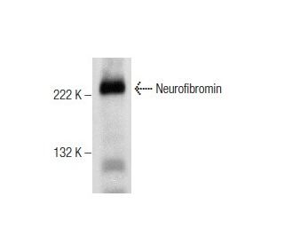
O Anticorpo Neurofibromina (H-12) é um anticorpo monoclonal IgG1 κ de camundongo contra Neurofibromina (também designado anticorpo NF1) que detecta a proteína Neurofibromina de origem camundongo, rato e humana por WB, IP, IF, IHC(P) e ELISA. O anticorpo Neurofibromin (H-12) está disponível na forma de anticorpo anti-Neurofibromin não conjugado, bem como em várias formas conjugadas de anticorpo anti-Neurofibromin, incluindo agarose, HRP, PE, FITC e vários conjugados Alexa Fluor®. A neurofibromatose tipo 1 (NF1), ou neurofibromatose de von Reckinghausen, é um dos distúrbios autossômicos dominantes mais comuns em humanos. A análise de ligação inicial mapeou o gene NF1 no cromossomo 17. O transcrito previsto do NF1 codifica a proteína Neurofibromina de 2.818 aminoácidos, também designada como proteína relacionada ao NF1-GAP (NF1GRP). Por meio da análise da sequência, foi demonstrada a semelhança entre uma pequena região da Neurofibromina e membros da família genética Ras GAP. Funcionalmente, a Neurofibromina foi demonstrada por análise bioquímica envolvendo a hidrólise de RAS-GAP e complementação funcional em levedura para se assemelhar ainda mais à proteína GAP. A proteína Neurofibromina é expressa em níveis relativamente constantes em uma ampla gama de linhagens celulares e tecidos, incluindo cérebro, pulmão, fígado, rim, baço, músculo e cólon. Embora pouco se saiba sobre a função da Neurofibromina, a homologia com o domínio catalítico de proteínas com atividade GTPase sugere que a Neurofibromina também pode interagir in vivo com as proteínas Ras.
Alexa Fluor® é uma marca comercial da Molecular Probes Inc., OR., EUA
LI-COR® e Odyssey® são marcas registadas da LI-COR Biosciences
Referencias do Neurofibromin Anticorpo (H-12):
- Sobre a degradação lisossómica da neurofibromina e a sua fosforilação em melanócitos em cultura. | Kaufmann, D., et al. 1999. Biol Chem. 380: 1071-8. PMID: 10543444
- Dissecção molecular de características isoladas da doença na neurofibromatose mosaica tipo 1. | Maertens, O., et al. 2007. Am J Hum Genet. 81: 243-51. PMID: 17668375
- Regulação do splicing alternativo específico dos neurónios do pré-mRNA da neurofibromatose tipo 1. | Zhu, H., et al. 2008. Mol Cell Biol. 28: 1240-51. PMID: 18086893
- Sinalização de RAS em carcinomas colorrectais através da alteração de RAS, RAF, NF1, e/ou RASSF1A. | Ahlquist, T., et al. 2008. Neoplasia. 10: 680-6, 2 p following 686. PMID: 18592002
- Identificação do produto do gene da neurofibromatose tipo 1. | Gutmann, DH., et al. 1991. Proc Natl Acad Sci U S A. 88: 9658-62. PMID: 1946382
- O gene da neurofibromatose tipo 1 codifica uma proteína relacionada com a GAP. | Xu, GF., et al. 1990. Cell. 62: 599-608. PMID: 2116237
- O domínio catalítico do produto do gene da neurofibromatose tipo 1 estimula a ras GTPase e complementa os mutantes ira de S. cerevisiae. | Xu, GF., et al. 1990. Cell. 63: 835-41. PMID: 2121369
- O domínio relacionado com GAP do produto do gene da neurofibromatose tipo 1 interage com ras p21. | Martin, GA., et al. 1990. Cell. 63: 843-9. PMID: 2121370
- A neurofibromina 1 é um miRNA alvo nos neurónios. | Paschou, M. and Doxakis, E. 2012. PLoS One. 7: e46773. PMID: 23056445
- Análise de ligação multiponto na neurofibromatose tipo I: uma colaboração internacional. | Goldgar, DE., et al. 1989. Am J Hum Genet. 44: 6-12. PMID: 2491784
- A neurofibromina regula os ataques epilépticos no modelo de epilepsia induzido por pilocarpina no rato. | Ren, M., et al. 2016. Mol Neurobiol. 53: 6069-6077. PMID: 26537900
- O fator de recrutamento da neurofibromina Spred1 liga-se ao domínio relacionado com o GAP sem afetar a inativação de Ras. | Dunzendorfer-Matt, T., et al. 2016. Proc Natl Acad Sci U S A. 113: 7497-502. PMID: 27313208
Informacoes sobre ordens
| Nome do Produto | Numero de Catalogo | UNID | Preco | Qde | FAVORITOS | |
Neurofibromin Anticorpo (H-12) | sc-376886 | 200 µg/ml | $322.00 | |||
Neurofibromin Anticorpo (H-12) AC | sc-376886 AC | 500 µg/ml, 25% agarose | $424.00 | |||
Neurofibromin Anticorpo (H-12) HRP | sc-376886 HRP | 200 µg/ml | $322.00 | |||
Neurofibromin Anticorpo (H-12) FITC | sc-376886 FITC | 200 µg/ml | $336.00 | |||
Neurofibromin Anticorpo (H-12) PE | sc-376886 PE | 200 µg/ml | $349.00 | |||
Neurofibromin Anticorpo (H-12) Alexa Fluor® 488 | sc-376886 AF488 | 200 µg/ml | $364.00 | |||
Neurofibromin Anticorpo (H-12) Alexa Fluor® 546 | sc-376886 AF546 | 200 µg/ml | $364.00 | |||
Neurofibromin Anticorpo (H-12) Alexa Fluor® 594 | sc-376886 AF594 | 200 µg/ml | $364.00 | |||
Neurofibromin Anticorpo (H-12) Alexa Fluor® 647 | sc-376886 AF647 | 200 µg/ml | $364.00 | |||
Neurofibromin Anticorpo (H-12) Alexa Fluor® 680 | sc-376886 AF680 | 200 µg/ml | $364.00 | |||
Neurofibromin Anticorpo (H-12) Alexa Fluor® 790 | sc-376886 AF790 | 200 µg/ml | $364.00 |