Date published: 2025-9-9

001 800-1338-3838

SCBT Portrait Logo
Seach Input

FAS-L 항체 (NOK-1): sc-19681

3.9(14)
리뷰 쓰기질문하기

데이터 시트
  • FAS-L 항체 NOK-1 는 마우스 monoclonal IgG1 κ FAS-L 항체, 37간행물에 인용, 이며 200 µg/ml으로 제공합니다.
  • L5178Y 마우스 T 림프구 세포를 재조합 human FAS-L로 발현시킨 것에 대해 제기됨
  • FAS-L 항체 (NOK-1)는 WB, IP, IF, IHC(P) and FCM으로 mouse, rat and human유래의 FAS-L 를 감지하는 데에 추천한다.; WB의 감지에는 권장되지 않습니다.
  • 항-FAS-L 항체(NOK-1)는 IP용 agarose와 결합되어 이용 가능하며, WB, IHC(P), ELISA용 HRP와 결합되어 이용 가능하며, IF, IHC(P), FCM용 phycoerythrin 또는 FITC와 결합되어 이용 가능합니다.
  • WB (RGB), IF, IHC(P) 와FCM, RGB fluorescent imaging systems, such as iBright™ FL1000, FluorChem™, Typhoon, Azure and other comparable systems에 사용가능한 Alexa Fluor® 488, Alexa Fluor® 546, Alexa Fluor® 594 or Alexa Fluor® 647결합제품도 있습니다.
  • WB (NIR), IF와FCM,Near-Infrared (NIR) detection systems, such as LI-COR®Odyssey®, iBright™ FL1000, FluorChem™, Typhoon, Azure and other comparable systems에 사용가능한 Alexa Fluor® 680 or Alexa Fluor® 790 결합제품도 있습니다.
  • FAS-L 작용을 차단하기 위해 아지드가 없는 제품도 제공, sc-19988 L, 200 µg/0.1 ml
  • FAS-L (NOK-1): sc-19681무료10 µg 샘플을 신청하려면 기술지원팀 (또는 로컬 디스트리뷰터)에 연락주세요.
  • m-IgG Fc BP-HRPm-IgG1 BP-HRP 는 FAS-L 항체 (NOK-1) 가 WB and IHC(P) 에 응용될 때 우선으로 사용하는 이차 항체 검사 시약입니다.이 시약은FAS-L 항체(NOK-1) 와 세트로 제공드립니다. (아래의 주문 정보를 참고해 주시길 바랍니다)

빠른 링크

더보기

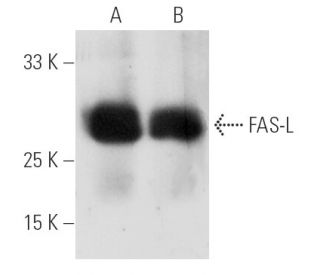
FAS-L 항체(NOK-1)는 마우스, 랫트, 그리고 인간 표본에서 웨스턴 블롯, 면역침전, 파라핀에 포함된 단면에서의 면역형광, 면역조직화학, 그리고 유세포분석을 통해 FAS-L 단백질(FASLG, CD178, CD95-L, TNFSF6, 또는 Fas 리간드라고도 함)을 검출하는 마우스 단일클론 IgG1 카파 경쇄 항체입니다. FAS-L 항체(NOK-1)는 비접합 형태와 아가로스, 양고추냉이 과산화효소, 피코에리트린, 플루오레세인 이소티오시아네이트, 여러 알렉사 플루오르® 접합체를 포함한 여러 접합 형태로 제공됩니다. 세포독성 T 림프구(CTL) 매개 세포독성은 바이러스에 감염된 세포나 변형된 세포에 대한 면역 감시에서 특정 효과기구의 중요한 구성요소를 구성합니다. 이 활동은 두 가지 메커니즘에 의해 설명되는데, 하나는 퍼포린 기반 과정입니다. FAS 기반 메커니즘은 전달 분자 FAS(Apo-1이라고도 함)와 FAS-L을 포함합니다. 인간 FAS 단백질은 CD40, 신경 성장 인자 수용체, 종양 괴사 인자 수용체를 포함하는 수용체 계열에 속하는 세포 표면 당단백질입니다. FAS 항원은 광범위한 림프구 세포주에서 발현되며, 그 중 일부는 항-FAS 항체 치료에 반응하여 세포 자멸사를 겪습니다. 이러한 발견은 표적 세포 사멸이 잠재적으로 FAS와 FAS-L 또는 이펙터의 세포 간 상호 작용에 의해 매개된다는 것을 강력하게 시사하며, FAS가 CTL 매개 세포 독성에 결정적으로 관여할 수 있음을 시사합니다.

연구용으로만 사용가능합니다. 진단이나 치료용으로 사용불가합니다.

Alexa Fluor®는 미국 오리건주 Molecular Probes Inc.의 상표입니다.

LI-COR®와 Odyssey®는 LI-COR Biosciences의 등록 상표입니다.

FAS-L 항체 (NOK-1) 참고문헌:

  1. 인간 미세아교세포에 대한 Fas-Ligand의 조절: 체외 연구.  |  Frigerio, S., et al. 2000. J Neuroimmunol. 105: 109-14. PMID: 10742551
  2. 인간 대장암 종양에서 Fas와 Fas-L 분자를 포함하는 종양 탈출 메커니즘.  |  Radfar, S., et al. 2000. Gastroenterol Clin Biol. 24: 1191-6. PMID: 11173732
  3. 고도로 악성 골수종 형질세포에 의한 Fas-L 상향 조절: 빈혈의 발병과 질병 진행에 있어서의 역할.  |  Silvestris, F., et al. 2001. Blood. 97: 1155-64. PMID: 11222356
  4. Fas/Fas 리간드 매개 세포 사멸에서 단백질 키나아제 C-테타의 중요한 역할.  |  Manicassamy, S. and Sun, Z. 2007. J Immunol. 178: 312-9. PMID: 17182568
  5. FasL과 TRAIL은 리슈마니아 메이저에 노출되면 표피 세포사멸과 피부 궤양을 유도합니다.  |  Eidsmo, L., et al. 2007. Am J Pathol. 170: 227-39. PMID: 17200196
  6. 만성 위염 및 십이지장 궤양 소아의 위 점막에서 가스트린, 소마토스타틴, PCNA 및 Fas-L의 발현 [만성 위염 및 십이지장 궤양].  |  Xie, XZ., et al. 2006. Zhonghua Er Ke Za Zhi. 44: 774-7. PMID: 17229384
  7. 세포 사멸의 매개체 Fas와 FasL은 다발성 경화증의 장애 진행을 10년 동안 예측합니다.  |  Lopatinskaya, L., et al. 2006. Mult Scler. 12: 704-9. PMID: 17262997
  8. FasL 단백질 전달을 통해 종양 내 CD4+CD25+ 조절 T 세포를 고갈시키면 입양 T 세포 전달의 치료 효능이 향상됩니다.  |  Chen, A., et al. 2007. Cancer Res. 67: 1291-8. PMID: 17283166
  9. [유육종증과 대조군에서 폐포 림프구의 세포 사멸은 비흡연자보다 흡연자에서 더 자주 발생합니다].  |  Kopiński, P., et al. 2006. Przegl Lek. 63: 841-7. PMID: 17288168
  10. 림프구 매개 세포 독성 메커니즘.  |  Henkart, PA. 1985. Annu Rev Immunol. 3: 31-58. PMID: 3904772
  11. T세포 매개 세포 독성의 일반적인 메커니즘에서 Fas와 그 리간드.  |  Hanabuchi, S., et al. 1994. Proc Natl Acad Sci U S A. 91: 4930-4. PMID: 7515183
  12. Fas 단백질은 정상 마우스의 CD4+CD8+ 흉선세포와 활성화된 성숙 림프구에서 높은 수준으로 발현되지만, 루푸스 취약 균주인 MRL lpr/lpr에서는 발현되지 않습니다.  |  Drappa, J., et al. 1993. Proc Natl Acad Sci U S A. 90: 10340-4. PMID: 7694292

주문정보

제품명카탈로그 번호 단위가격수량관심품목

FAS-L 항체 (NOK-1)

sc-19681
200 µg/ml
$316.00

FAS-L (NOK-1): m-IgG Fc BP-HRP 번들

sc-526589
200 µg Ab; 10 µg BP
$354.00

FAS-L (NOK-1): m-IgG1 BP-HRP 번들

sc-531962
200 µg Ab; 20 µg BP
$354.00

FAS-L 항체 (NOK-1) L

sc-19681 L
200 µg/0.1 ml
$316.00

FAS-L 항체 (NOK-1) AC

sc-19681 AC
500 µg/ml, 25% agarose
$416.00

FAS-L 항체 (NOK-1) HRP

sc-19681 HRP
200 µg/ml
$316.00

FAS-L 항체 (NOK-1) FITC

sc-19681 FITC
200 µg/ml
$330.00

FAS-L 항체 (NOK-1) PE

sc-19681 PE
200 µg/ml
$343.00

FAS-L 항체 (NOK-1) Alexa Fluor® 488

sc-19681 AF488
200 µg/ml
$357.00

FAS-L 항체 (NOK-1) Alexa Fluor® 546

sc-19681 AF546
200 µg/ml
$357.00

FAS-L 항체 (NOK-1) Alexa Fluor® 594

sc-19681 AF594
200 µg/ml
$357.00

FAS-L 항체 (NOK-1) Alexa Fluor® 647

sc-19681 AF647
200 µg/ml
$357.00

FAS-L 항체 (NOK-1) Alexa Fluor® 680

sc-19681 AF680
200 µg/ml
$357.00

FAS-L 항체 (NOK-1) Alexa Fluor® 790

sc-19681 AF790
200 µg/ml
$357.00

I am looking to utilize this antibody to neutralize FAS-L. Do I need to specify that I need it azide-free?

Asked by: Jame Dolan
Thank you for your question. To request an azide-free antibody please contact our special orders department for a price quote and lead time. Please email specialorders@scbt.com
Answered by: BlakeJ
Date published: 2023-05-18

This particular antibody is listed as not for use with western blot, but the majority of the reviews are for western blots including a review from your QC department. What's really going on with this antibody?

Asked by: Allyson
Thank you for your question. We do not recommend this mouse monoclonal antibody, FAS-L (NOK-1): sc-19681, for use in Western blotting. Based on our in-house testing and data on the website the antibody is recommended for IP, IF, IHC(P) and FCM. We will remove the review from our QC department which mistakenly interpreted our published IP data with the antibody as WB data (because the WB was performed with a different antibody).
Answered by: Technical Support
Date published: 2017-11-22

Hello, out of Fas-Ligand antibodies N-20, C-178, Q-20, and C-20, only C-20 has given me consistent results in western blotting. NOK-1 antibody, the suggested replacement for C-20, is not listed for western blotting. Which Fas-L antibody should I use?

Asked by: Nayan
Thank you for your question. The NOK-1 clone is a well established antibody against FAS-L that is commonly used for flow cytometry (FCM), IF, IHC(P) and immunoprecipitation. It is also available azide-free for neutralizing the cytotoxic effects of FAS-L. This clone is not recommended for Western blotting. We do offer alternative clones that are specifically recommended for Western blotting. For human samples, we recommend, FAS-L Antibody (2.1): sc-33716. And for mouse samples we recommend, FAS-L Antibody (Kay-10): sc-19988. Links to these product pages are provided below. https://www.scbt.com/scbt/product/fas-l-antibody-2-1 https://www.scbt.com/scbt/product/fas-l-antibody-kay-10
Answered by: Technical Support
Date published: 2017-03-13

Can FAS-L (NOK-1): sc-19681 monoclonal antibody be used in IF or IHC with mouse tissue or cells? Will there be non-specific staining?

Asked by: AbPolly
Thank you for your inquiry. The use of mouse monoclonal antibodies with mouse samples is very common and typically poses no problem. To eliminate any potential cross-reactivity of an anti-mouse secondary detection reagent, FAS-L (NOK-1): sc-19681 is available in a variety of direct conjugations, such as HRP, PE, FITC and AlexaFluors.
Answered by: Technical Support
Date published: 2017-02-24
  • y_2025, m_9, d_5, h_6CST
  • bvseo_bulk, prod_bvqa, vn_bulk_3.0.42
  • cp_1, bvpage1
  • co_hasquestionsanswers, tq_4
  • loc_ko_KR, sid_19681, prod, sort_[SortEntry(order=LAST_APPROVED_ANSWER_SUBMISSION_TIME, direction=DESCENDING)]
  • clientName_scbt
  • bvseo_sdk, java_sdk, bvseo-4.0.0
  • CLOUD, getContent, 116ms
  • QUESTIONS, PRODUCT
Rated 5 out of 5 by from Quite good antibody!1:200 diluted for wb of rat protein ,the band is strong and specia!
Date published: 2017-12-30
Rated 5 out of 5 by from WBI have used this FAS-L antibody for many times to verify the expression in mosue kidney tissue .I Never meet problems of background,it always shows strong signal for WB use 1:1000.
Date published: 2017-08-18
Rated 5 out of 5 by from clear signal bandI'm running western blots for FAS-L protein analysis,In my experience,this antibody can diluted at 1:500 to 1:1000. 1:200 is too much background.The loading amount and incubated conditions should be optimitized.
Date published: 2017-07-22
Rated 5 out of 5 by from Clean bandClean band observed in Western Blot analysis of FAS-L expression in HeLa whole cell lysate
Date published: 2017-05-24
Rated 5 out of 5 by from Worked goodThis antibody work well in the western blot with the dilution 1:800.
Date published: 2017-05-13
Rated 5 out of 5 by from Works greatWorks great for paraffin embedded human lung tissue sections,I suggest use dilution 1:300 for IF.
Date published: 2017-05-13
Rated 3 out of 5 by from Decent immunoperoxidase cytoplasmic stainingDecent immunoperoxidase cytoplasmic staining in formalin fixed, paraffin-embedded human pancreas tissue. -SCBT QC
Date published: 2015-05-13
Rated 3 out of 5 by from Published data for IP in human Ramos cellsPublished data for IP in human Ramos cells. -SCBT Publication Review
Date published: 2015-04-22
  • y_2025, m_9, d_5, h_6
  • bvseo_bulk, prod_bvrr, vn_bulk_3.0.42
  • cp_1, bvpage1
  • co_hasreviews, tv_1, tr_13
  • loc_ko_KR, sid_19681, prod, sort_[SortEntry(order=SUBMISSION_TIME, direction=DESCENDING)]
  • clientName_scbt
  • bvseo_sdk, java_sdk, bvseo-4.0.0
  • CLOUD, getReviews, 16ms
  • REVIEWS, PRODUCT
FAS-L 항체 (NOK-1) is rated 3.9 out of 5 by 14.
  • y_2025, m_9, d_5, h_6
  • bvseo_bulk, prod_bvrr, vn_bulk_3.0.42
  • cp_1, bvpage1
  • co_hasreviews, tv_1, tr_13
  • loc_ko_KR, sid_19681, prod, sort_[SortEntry(order=SUBMISSION_TIME, direction=DESCENDING)]
  • clientName_scbt
  • bvseo_sdk, java_sdk, bvseo-4.0.0
  • CLOUD, getAggregateRating, 118ms
  • REVIEWS, PRODUCT