")
EphB3 Anticorpo (7E5): sc-100299
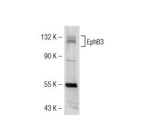
- EphB3 Anticorpo 7E5é um anticorpo monoclonal produzido em camundongo IgG1 κ, citado em 4 publicações, fornecido em 100 µg/ml
- Produzido contra recombinante EphB3 de human origem
- recomendado para a detecção de EphB3 de mouse, rat e human origem em
- Atualmente, ainda não concluímos a identificação do(s) reagente(s) de deteção secundário(s) preferido(s) para EphB3 Anticorpo (7E5). Este trabalho está em curso.

A subfamília Eph representa o maior grupo de receptores de proteínas tirosina quinases identificado até à data. Embora as actividades biológicas destes receptores ainda não tenham sido determinadas, há cada vez mais provas de que estão envolvidos na função do sistema nervoso central e no desenvolvimento. Os receptores da subfamília Eph de origem humana (e os seus homólogos murinos/avianos) incluem EphA1 (Eph), EphA2 (Eck), EphA3 (Hek4), EphA4 (Hek8), EphA5 (Hek7), EphA6 (Hek12), EphA7 (Hek11/MDK1), EphA8 (Hek3), EphB1 (Hek6), EphB2 (Hek5), EphB3 (Cek10, Hek2), EphB4 (Htk), EphB5 (Hek9) e EphB6 (Mep). Os ligandos dos receptores Eph incluem a efrina-A4 (LERK-4), que se liga ao EphA3 e ao EphB1. Além disso, a efrina-A2 (ELF-1) foi descrita como ligando o EphA4, a efrina-A3 (Ehk1-L) como ligando o EphA5 e a efrina-B2 (Htk-L) como ligando o EphB4 (Htk).
Alexa Fluor® é uma marca comercial da Molecular Probes Inc., OR., EUA
LI-COR® e Odyssey® são marcas registadas da LI-COR Biosciences
Referencias do EphB3 Anticorpo (7E5):
- EphB3 marca a delaminação das células progenitoras endócrinas no pâncreas em desenvolvimento. | Villasenor, A., et al. 2012. Dev Dyn. 241: 1008-19. PMID: 22434763
- Papel do Recetor EphB3 na Mediação do Crescimento do Tumor da Cabeça e Pescoço, Migração Celular e Resposta ao Inibidor PI3K. | Bhatia, S., et al. 2018. Mol Cancer Ther. 17: 2049-2059. PMID: 29970482
- Recetor tirosina quinase EphB3: um indicador de prognóstico no carcinoma colorrectal. | Xuan, Z., et al. 2020. Pathol Oncol Res. 26: 541-549. PMID: 30535864
- Significado prognóstico e clinicopatológico da expressão de EphB3 e Disaderina no colangiocarcinoma extra-hepático. | Wu, Z., et al. 2020. Cancer Manag Res. 12: 221-232. PMID: 32021438
- Perfil de Expressão e Significado Prognóstico do EPHB3 no Cancro Colorrectal. | Jang, BG., et al. 2020. Biomolecules. 10: PMID: 32294981
- EphB3 como um potencial mediador da osteogénese reparadora e do desenvolvimento. | Kamath, RAD. and Benson, MD. 2023. Cells Tissues Organs. 212: 125-137. PMID: 34695818
- A redução do EphB3 inibe a proliferação celular, em parte através da via de sinalização AKT e reprime a transição epitelial-mesenquimal no carcinoma de células escamosas do esófago. | Tang, L., et al. 2022. Transl Cancer Res. 11: 85-98. PMID: 35261887
- Papel e mecanismo da EphB3 nas crises epilépticas e na epileptogénese através da Kalirina. | Huang, H., et al. 2024. Mol Cell Neurosci. 128: 103915. PMID: 38143048
- O recetor EphB3 suprime a invasão, migração e proliferação em gliomas através da inibição da via de sinalização EGFR-PI3K/AKT. | Xiao, Z., et al. 2024. Brain Res. 1830: 148796. PMID: 38341169
- A ausência do recetor EphB3 previne a perda óssea em modelos de osteoporose em ratos. | Rodríguez-Sosa, MR., et al. 2024. J Bone Miner Res. 39: 1008-1024. PMID: 38739682
Informacoes sobre ordens
| Nome do Produto | Numero de Catalogo | UNID | Preco | Qde | FAVORITOS | |
EphB3 Anticorpo (7E5) | sc-100299 | 100 µg/ml | $339.00 |