Date published: 2025-9-9

001 800-1338-3838

SCBT Portrait Logo
Seach Input

APC 항체 (F-3): sc-9998

4.4(20)
리뷰 쓰기질문하기

데이터 시트
  • APC 항체 F-3 는 마우스 monoclonal IgG1 κ APC 항체, 33간행물에 인용, 이며 200 µg/ml으로 제공합니다.
  • 아미노 산2-289 mapping at the N-terminus of APC of human 의하고 기원한다
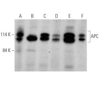
  • APC 항체 (F-3)는 WB, IP, IF, IHC(P) and ELISA으로 mouse, rat and human유래의 APC 를 감지하는 데에 추천한다.
  • 항-APC 항체(F-3)는 IP용 agarose와 결합되어 이용 가능하며, WB, IHC(P), ELISA용 HRP와 결합되어 이용 가능하며, IF, IHC(P), FCM용 phycoerythrin 또는 FITC와 결합되어 이용 가능합니다.
  • WB (RGB), IF, IHC(P) 와FCM, RGB fluorescent imaging systems, such as iBright™ FL1000, FluorChem™, Typhoon, Azure and other comparable systems에 사용가능한 Alexa Fluor® 488, Alexa Fluor® 546, Alexa Fluor® 594 or Alexa Fluor® 647결합제품도 있습니다.
  • WB (NIR), IF와FCM,Near-Infrared (NIR) detection systems, such as LI-COR®Odyssey®, iBright™ FL1000, FluorChem™, Typhoon, Azure and other comparable systems에 사용가능한 Alexa Fluor® 680 or Alexa Fluor® 790 결합제품도 있습니다.
  • APC (F-3): sc-9998무료10 µg 샘플을 신청하려면 기술지원팀 (또는 로컬 디스트리뷰터)에 연락주세요.
  • m-IgG Fc BP-HRPm-IgG1 BP-HRP 는 APC 항체 (F-3) 가 WB and IHC(P) 에 응용될 때 우선으로 사용하는 이차 항체 검사 시약입니다.이 시약은APC 항체(F-3) 와 세트로 제공드립니다. (아래의 주문 정보를 참고해 주시길 바랍니다)

빠른 링크

더보기

APC 항체(F-3)는 마우스, 쥐, 인간에서 유래된 대장용종성 대장염(APC) 단백질을 웨스턴 블랏팅, 면역침전, 면역형광, 면역조직화학, 효소결합면역흡착분석 등 다양한 응용을 통해 검출하는 마우스 단일클론 IgG1 카파 경쇄 항체입니다. APC 단일 클론 항체(F-3)는 비접합 형태와 아가로스, 호스래디시 퍼옥시다아제, 피코에리트린, 플루오레세인 이소티오시아네이트, 다양한 알렉사 플루오르® 접합체 등 여러 가지 접합체 형태로 제공됩니다. APC 단백질은 세포 부착을 조절하고 상피 조직의 완전성을 유지하는 데 중요한 역할을 하는데, 이는 종양 발생을 예방하는 데 필수적입니다. APC 유전자의 돌연변이는 일반적으로 가족성 선종성 용종증과 가드너 증후군과 관련이 있는데, 이 두 질환은 결장암으로 이어질 수 있는 수많은 선종성 용종의 발생을 특징으로 합니다. 2,843개의 아미노산으로 구성된 APC 단백질은 어디에나 존재하며, 세포 부착에 필수적인 β-카테닌과 γ-카테닌을 포함한 세포 내 접합부의 주요 구조적 구성 요소와 상호 작용합니다. APC는 β- 및 γ-catenin의 특정 영역에 결합하기 위해 E-cadherin과 경쟁하며, 세포 골격과 APC-E-cadherin-β/γ-catenin 복합체 사이의 상호 작용은 α-catenin에 의해 매개되어 세포 성장과 분화를 조절하는 세포 신호 전달 경로에서 APC의 중요성을 강조합니다.

연구용으로만 사용가능합니다. 진단이나 치료용으로 사용불가합니다.

Alexa Fluor®는 미국 오리건주 Molecular Probes Inc.의 상표입니다.

LI-COR®와 Odyssey®는 LI-COR Biosciences의 등록 상표입니다.

APC 항체 (F-3) 참고문헌:

  1. 대장암의 APC 유전자.  |  Fodde, R. 2002. Eur J Cancer. 38: 867-71. PMID: 11978510
  2. FAP 및 대장암 환자에서 염색체 5q21 유전자의 돌연변이.  |  Nishisho, I., et al. 1991. Science. 253: 665-9. PMID: 1651563
  3. APC와 발암성 KRAS는 장 종양 형성 및 진행에서 Wnt 신호를 강화하는 데 시너지 효과를 발휘합니다.  |  Janssen, KP., et al. 2006. Gastroenterology. 131: 1096-109. PMID: 17030180
  4. Myc 결실은 소장에서 APC 결핍을 구제합니다.  |  Sansom, OJ., et al. 2007. Nature. 446: 676-9. PMID: 17377531
  5. FAP 관련 갑상선 암종 발생에서 APC 유전자 및/또는 ss 카테닌 유전자의 생식선 및 체세포 돌연변이.  |  Cetta, F., et al. 2007. World J Surg. 31: 1366-7; author reply 1368-9. PMID: 17426899
  6. B-카테닌 결핍은 Myc 결핍이 아닌 간에서 APC 손실의 즉각적인 표현형 발현을 억제합니다.  |  Reed, KR., et al. 2008. Proc Natl Acad Sci U S A. 105: 18919-23. PMID: 19033191
  7. 기계적 요인이 APC 마우스 결장에서 베타카테닌 의존성 종양 유전자 발현을 활성화합니다.  |  Whitehead, J., et al. 2008. HFSP J. 2: 286-94. PMID: 19404440
  8. Wnt 신호의 Apc 억제는 배아 발생 과정과 성인기 동안의 상아 치아 형성을 조절합니다.  |  Wang, XP., et al. 2009. Development. 136: 1939-49. PMID: 19429790
  9. 발달 중인 뇌의 APC/C와 CK1.  |  Penas, C., et al. 2015. Oncotarget. 6: 16792-3. PMID: 26219466
  10. 위장암에서 APC 프로모터 메틸화.  |  Zhu, L., et al. 2021. Front Oncol. 11: 653222. PMID: 33968756
  11. METTL3은 APC mRNA N6-메틸아데노신 의존성 YTHDF 결합에 의해 매개되는 APC 발현을 감소시킴으로써 종양 발생을 촉진합니다.  |  Wang, W., et al. 2021. Nat Commun. 12: 3803. PMID: 34155197
  12. 선종성 용종증 대장균 유전자(APC) 영역의 고해상도 유전자 지도.  |  Olschwang, S., et al. 1995. Am J Med Genet. 56: 413-9. PMID: 7604852

주문정보

제품명카탈로그 번호 단위가격수량관심품목

APC 항체 (F-3)

sc-9998
200 µg/ml
$316.00

APC (F-3): m-IgG Fc BP-HRP 번들

sc-526532
200 µg Ab; 10 µg BP
$354.00

APC (F-3): m-IgG1 BP-HRP 번들

sc-531905
200 µg Ab; 20 µg BP
$354.00

APC 항체 (F-3) AC

sc-9998 AC
500 µg/ml, 25% agarose
$416.00

APC 항체 (F-3) HRP

sc-9998 HRP
200 µg/ml
$316.00

APC 항체 (F-3) FITC

sc-9998 FITC
200 µg/ml
$330.00

APC 항체 (F-3) PE

sc-9998 PE
200 µg/ml
$343.00

APC 항체 (F-3) Alexa Fluor® 488

sc-9998 AF488
200 µg/ml
$357.00

APC 항체 (F-3) Alexa Fluor® 546

sc-9998 AF546
200 µg/ml
$357.00

APC 항체 (F-3) Alexa Fluor® 594

sc-9998 AF594
200 µg/ml
$357.00

APC 항체 (F-3) Alexa Fluor® 647

sc-9998 AF647
200 µg/ml
$357.00

APC 항체 (F-3) Alexa Fluor® 680

sc-9998 AF680
200 µg/ml
$357.00

APC 항체 (F-3) Alexa Fluor® 790

sc-9998 AF790
200 µg/ml
$357.00

The full length of APC is 310kD, why there are two bands around 100kD? and why do we see in some blots two bands around 100kD and in others one 310kD band? In my western i got the two bands around 100kD and no band around 310kD, is it APC?

Asked by: CaGI
Thank you for your question. Please contact our Technical Service Department or your local distributor for assistance in troubleshooting this antibody.
Answered by: Tech Service 1
Date published: 2020-12-07

I am using this antibody for APC detection (SC-9998). I am detecting the full lenght form which is about 311 KD, but, I see 2 bands instead only 1 in this place. My question is Why I see 2 bandas around 312 KD?? also, I detect a band around 140 KD. Why?

Asked by: ali jose calderon
Thank you for your question. We have not seen doublet bands in the higher band size, but as this protein can be ubiquitinated that could be a possible reason. It is also sometimes due to the percentage gel used. If you have any further questions, please contact Technical Service by phone, (800)-457-3801 option 2, email <scbt@scbt.com>, or by live chat directly on our website, www.scbt.com.
Answered by: Tech Service
Date published: 2017-05-26

Do you have any tips for performing a double stain with only mouse monoclonal antibodies?

Asked by: Cweed
We recommend using directly conjugated primary antibodies. This product, sc-9998, is available conjugated to FITC, AF488, and AF647.
Answered by: Technical Support 12
Date published: 2017-01-12

Will this antibody work in canine samples?

Asked by: Cweed
Please contact our Technical Service Department and we would be happy to perform sequence analysis and review our data to answer any questions you may have about additional species reactivity.
Answered by: Technical Support 12
Date published: 2017-01-12
  • y_2025, m_8, d_28, h_5CST
  • bvseo_bulk, prod_bvqa, vn_bulk_3.0.42
  • cp_1, bvpage1
  • co_hasquestionsanswers, tq_4
  • loc_ko_KR, sid_9998, prod, sort_[SortEntry(order=LAST_APPROVED_ANSWER_SUBMISSION_TIME, direction=DESCENDING)]
  • clientName_scbt
  • bvseo_sdk, java_sdk, bvseo-4.0.0
  • CLOUD, getContent, 111ms
  • QUESTIONS, PRODUCT
Rated 5 out of 5 by from good performanceThis antibody works well in Western blotting in our knockdown experiment
Date published: 2022-05-27
Rated 5 out of 5 by from Good for IFThis APC antibody shows strong specific signal in IF staining in primary rat motor neuron.
Date published: 2017-11-06
Rated 4 out of 5 by from good antibody and works well in western blot.tested protein extracts from murine organoids with genetic as well as Cripsr/Cas9 mediated deletion of APC
Date published: 2017-10-19
Rated 1 out of 5 by from Not specific on HCT-116 cell lineI prove it on HCT-116 cell line and I observed multiple bands but neither of them correspond to APC.
Date published: 2017-09-29
Rated 5 out of 5 by from WB good result2 bands can be detected around 310kD and 130kD in Caco2 cell lines,I wonder why? I dilute it in 5% dry skim milk,1:200, and overnight in 4℃.
Date published: 2017-09-26
Rated 1 out of 5 by from No bandNo band on WB. Used 1:1000 dilution for MCF-7 cell lysates.
Date published: 2017-07-03
Rated 3 out of 5 by from Unfortunately not for IFI tested this antibody on human endothelial HUVEC cells as well as on mouse oocytes but unfortunately the antibody did not work in these immunofluorescence experiments. I always had alpha-tubulin as positive control, which always worked well.
Date published: 2017-06-22
Rated 4 out of 5 by from performance in HCC cell lineI tested the sample APC antibody in Huh7 and SK-Hep1 cell lines. Both cells were untreated or transfected with pEGFP-N1/ overexpressing plasmid. In all the groups, western blot result (Bio-Rad Clarity Western ECL) showed one stripe above the 100kDa marker. But I notice that this APC antibody could also recognize GFP tag, two different pEGFP-N1/ overexpressing plasmids with different protein size could be showen in the western blot result and they were way brighter than the suppose APC. It could be disturbing for some experiments.
Date published: 2017-06-07
  • y_2025, m_8, d_28, h_5
  • bvseo_bulk, prod_bvrr, vn_bulk_3.0.42
  • cp_1, bvpage1
  • co_hasreviews, tv_1, tr_19
  • loc_ko_KR, sid_9998, prod, sort_[SortEntry(order=SUBMISSION_TIME, direction=DESCENDING)]
  • clientName_scbt
  • bvseo_sdk, java_sdk, bvseo-4.0.0
  • CLOUD, getReviews, 18ms
  • REVIEWS, PRODUCT
APC 항체 (F-3) is rated 4.4 out of 5 by 20.
  • y_2025, m_8, d_28, h_5
  • bvseo_bulk, prod_bvrr, vn_bulk_3.0.42
  • cp_1, bvpage1
  • co_hasreviews, tv_1, tr_19
  • loc_ko_KR, sid_9998, prod, sort_[SortEntry(order=SUBMISSION_TIME, direction=DESCENDING)]
  • clientName_scbt
  • bvseo_sdk, java_sdk, bvseo-4.0.0
  • CLOUD, getAggregateRating, 157ms
  • REVIEWS, PRODUCT