")
: sc-56878. A549(A), MCF7(B), HeLa(C), MIA PaCa-2(D), ZR-75-1(E) 및 BT-20(F) 전체 세포 용해물에서 Akt1/2/3 발현의 웨스턴 블롯 분석.")
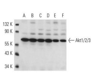
Akt1/2/3 항체(BDI111): sc-56878. A549(A), MCF7(B), HeLa(C), MIA PaCa-2(D), ZR-75-1(E) 및 BT-20(F) 전체 세포 용해물에서 Akt1/2/3 발현의 웨스턴 블롯 분석.
Akt1/2/3 항체 (BDI111): sc-56878
- Akt1/2/3 항체 BDI111 는 마우스 monoclonal IgG1, 60간행물에 인용, 이며 50 µg/0.5 ml으로 제공합니다.
- human의 Akt1의 아미노산 에 해당하는 합성 펩티드에 대해 제기됨
- WB 와IP으로 human origin의 Akt1 when it is dephosphorylated at Ser 473, Akt2 when it is dephosphorylated at Ser 474 and Akt3 when it is dephosphorylated at Ser 472을 검출할것을 권장합니다.
- See Akt1 (B-1): sc-5298 for additional antibody conjugates, including AC, HRP, FITC, PE, Alexa Fluor® 488, 594, 647, 680 and 790.
- 현재 Akt1/2/3 항체(BDI111)에 대해 선호하는 2차 검출 시약의 식별을 아직 완료하지 못했습니다. 이 작업은 진행 중입니다.

세린/트레오닌 키나제 Akt 계열은 단백질 키나제 A 및 C 계열과 서열 상동성을 나타내고 c-Akt 프로토-온코제인에 의해 암호화되는 Akt1(PKB 또는 RacPK), Akt2(PKB γ 또는 흉선 바이러스 프로토-온코제인 3으로도 지정됨)을 포함한 여러 구성원을 포함한다. Akt 계열의 모든 구성원은 플렉스스트린 상동성 도메인을 가지고 있다. Akt1 및 Akt2는 PDGF 자극에 의해 활성화된다. 활성화는 포스파티딜이노시톨 3-키나제(PI 3-키나제) 복합체의 하위 유닛에 결합하는 PDGFR-β Tyr 잔기 740 및 751에 의존한다. 인슐린 또는 인슐린 성장인자-1(IGF-1)에 의한 Akt1의 활성화는 Thr 308 및 Ser 473의 인산화를 초래한다. 두 잔기의 인산화는 높은 수준의 Akt1 활성을 생성하는 데 중요하다. Thr 308의 인산화는 생체 내에서 Ser 473의 인산화에 의존하지 않는다. 따라서, Akt 단백질은 상류 키나제(들)에 의해 인슐린/IGF-1 자극 세포에서 인산화되고 활성화된다. Akt1 및 Akt2의 활성화는 PI 키나제 억제제 wortmanin에 의해 억제되며, 이는 단백질이 PI 키나제의 하류에서 신호를 보낸다는 것을 시사한다.
연구용으로만 사용가능합니다. 진단이나 치료용으로 사용불가합니다.
Alexa Fluor®는 미국 오리건주 Molecular Probes Inc.의 상표입니다.
LI-COR®와 Odyssey®는 LI-COR Biosciences의 등록 상표입니다.
Akt1/2/3 항체 (BDI111) 참고문헌:
- 소라페닙은 인간 신경모세포종 세포주에서 ERK/Akt 및 STAT3 생존 경로를 하향 조절하고 세포 사멸을 유도합니다. | Chai, H., et al. 2010. Int J Clin Exp Pathol. 3: 408-15. PMID: 20490331
- 인간 요로상피의 증식은 비정형 β1 -아드레날린 수용체에 의해 유도됩니다. | Winder, M., et al. 2015. Auton Autacoid Pharmacol. 35: 32-40. PMID: 26913580
- 마우스 흑색종 세포에서 플라보노이드 유도체인 모린-7-황산나트륨의 항암 효과. | Li, HW., et al. 2016. Biomed Pharmacother. 84: 909-916. PMID: 27764752
- 인터루킨 6 결핍이 젊은 쥐와 늙은 쥐에서 박테리아 지질 다당류에 의해 유도된 심근 신호 전달 경로 활성화에 미치는 영향. | Tarasiuk, E., et al. 2020. Adv Med Sci. 65: 386-393. PMID: 32693349
- 세툭시맙 내성 두경부 편평세포암종에서 Akt의 역할: 새로운 병용 전략에 대한 시험관 내 연구. | Zaryouh, H., et al. 2021. Front Oncol. 11: 697967. PMID: 34568028
- 비소세포폐암에 대한 CDK4 억제제 파스카플라신과 EGFR 억제제 아파티닙의 세포 독성 시너지 효과. | Plangger, A., et al. 2022. Invest New Drugs. 40: 215-223. PMID: 34596822
- 지방세포와 아디포카인은 자궁 평활근종에 우호적인 미세 환경을 촉진합니다. | Afrin, S., et al. 2023. Nutrients. 15: PMID: 36771423
- H2S는 심장 β-아드레날린 수용체의 하류에서 G6PD 의존적인 방식으로 산화 환원 신호를 조절합니다. | Chhabra, A., et al. 2023. Cell Signal. 107: 110664. PMID: 37004833
- 생식력, 발정 주기 및 자궁 내막 발달에서 AKT1과 AKT2의 중복된 역할. | Adam, P., et al. 2023. Reproduction. 165: 605-616. PMID: 37053038
- AKT1은 헥소키나아제를 직접 인산화하여 종양 형성과 전이를 촉진합니다. | Yu, Y., et al. 2024. J Cell Biochem. 125: e30613. PMID: 38860522
주문정보
| 제품명 | 카탈로그 번호 | 단위 | 가격 | 수량 | 관심품목 | |
Akt1/2/3 항체 (BDI111) | sc-56878 | 50 µg/0.5 ml | $322.00 |