")
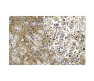
: sc-25350. Coloração por imunoperoxidase de tecido do pâncreas humano fixado em formalina e incluído em parafina, mostrando coloração citoplasmática do pâncreas exócrino e das células das ilhotas. Gentilmente cedido pelo programa sueco Human Protein Atlas (HPA).")
: sc-25350. Coloração por imunofluorescência de células K-562 fixadas em metanol mostrando a localização citoplasmática.")
: sc-25350. Análise de Western blot da proteína de fusão Sos 2 recombinante humana.")
Sos 2 Anticorpo (G-7): sc-25350
- Sos 2 Anticorpo G-7é um anticorpo monoclonal produzido em camundongo IgG2b (kappa light chain) fornecido em 200 µg/ml
- Produzido contra aminoácidos 1091-1170 de Sos 2 de human origem
- recomendado para a detecção de Sos 2 of human origin by WB, IP, IF, IHC(P) and ELISA
- m-IgG Fc BP-HRP, 2b BP-HRP">m-IgG2b BP-HRP e m-IgGκ BP-HRP são os reagentes de deteção secundários preferidos para Sos 2 Antibody (G-7) para aplicações WB e IHC(P). Estes reagentes são agora oferecidos em pacotes com Sos 2 Antibody (G-7)(ver informações de encomenda abaixo).

LINKS RÁPIDOS
A superfamília de proteínas de ligação a GTP, da qual as proteínas Ras são protótipos, tem sido implicada numa vasta gama de actividades biológicas. Estudos identificaram uma família de factores de libertação de nucleótidos de guanina (GRF) que activam Ras em células de mamíferos e uma proteína "adaptadora" (Sem 5/GRB2) que parece mediar a interação dos GRF com moléculas receptoras activadas. A Ras-GRF p140 promove a troca de nucleótidos na Ras p21s, mas não noutros membros da superfamília do gene Ras. Além disso, foram descritos três homólogos em mamíferos do Ras-GRF de Drosophila, son of sevenless (Sos). Estes incluem dois de camundongo, mSos 1 e mSos 2, e um de humano, hSos. Foi relatado que Vav p95 funciona como um GRF na ativação de Ras pelo recetor de células T e que tem um domínio semelhante ao de Dbl p115, que é um GRF específico para Cdc42Hs. Após a ativação, Ras parece interagir com Raf, activando assim a via de fosforilação da MAP quinase.
Alexa Fluor® é uma marca comercial da Molecular Probes Inc., OR., EUA
LI-COR® e Odyssey® são marcas registadas da LI-COR Biosciences
Referencias do Sos 2 Anticorpo (G-7):
- As variantes raras em SOS2 e LZTR1 estão associadas à síndrome de Noonan. | Yamamoto, GL., et al. 2015. J Med Genet. 52: 413-21. PMID: 25795793
- Mutações activadoras que afectam o domínio de homologia Dbl do SOS2 causam a síndrome de Noonan. | Cordeddu, V., et al. 2015. Hum Mutat. 36: 1080-7. PMID: 26173643
- As isoformas RAS oncogénicas apresentam um requisito hierárquico para o fator de troca de nucleótidos de guanina SOS2 para mediar a transformação celular. | Sheffels, E., et al. 2018. Sci Signal. 11: PMID: 30181243
- Condições de crescimento independentes de ancoragem revelam uma dependência diferencial de SOS2 para a transformação e sobrevivência em células cancerígenas com mutações RAS. | Sheffels, E., et al. 2021. Small GTPases. 12: 67-78. PMID: 31062644
- As variantes do gene SOS2 são uma causa rara da síndrome de Noonan com uma predisposição especial para complicações linfáticas. | Lissewski, C., et al. 2021. Eur J Hum Genet. 29: 51-60. PMID: 32788663
- SOS2 vem à tona: Differential Functionalities in Physiology and Pathology (Funcionalidades Diferenciais em Fisiologia e Patologia). | Baltanás, FC., et al. 2021. Int J Mol Sci. 22: PMID: 34205562
- As variantes genéticas de SOS2, MAP2K1 e RASGRF2 nos genes da via RAS prevêem a sobrevivência de doentes com carcinoma hepatocelular relacionado com o VHB. | Lin, Q., et al. 2023. Arch Toxicol. 97: 1599-1611. PMID: 37029817
- SOS2 modula o limiar da sinalização EGFR para regular a eficácia e a resistência do osimertinib no adenocarcinoma do pulmão. | Theard, PL., et al. 2024. Mol Oncol. 18: 641-661. PMID: 38073064
- Descoberta de pequenas moléculas que se ligam ao Son of Sevenless 2 (SOS2). | Zak, KM., et al. 2025. J Med Chem. 68: 2680-2693. PMID: 39818983
- Interações diferenciais de Sos1 e Sos2 humanas com Grb2. | Yang, SS., et al. 1995. J Biol Chem. 270: 18212-5. PMID: 7629138
Informacoes sobre ordens
| Nome do Produto | Numero de Catalogo | UNID | Preco | Qde | FAVORITOS | |
Sos 2 Anticorpo (G-7) | sc-25350 | 200 µg/ml | $322.00 | |||
Pacote do Sos 2 (G-7): m-IgG Fc BP-HRP | sc-536707 | 200 µg Ab; 10 µg BP | $361.00 | |||
Pacote do Sos 2 (G-7): m-IgGκ BP-HRP | sc-533867 | 200 µg Ab; 40 µg BP | $361.00 | |||
Pacote do Sos 2 (G-7): m-IgG2b BP-HRP | sc-549821 | 200 µg Ab; 10 µg BP | $361.00 |