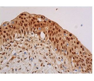
Date published: 2025-9-7

00800 4573 8000

SCBT Portrait Logo
Seach Input

Anticorpo p-Akt1/2/3 (B-12): sc-377556

4.2(10)
Scrivi una recensioneFai una domanda

Schede Tecniche
  • p-Akt1/2/3 Antibody B-12 è un monoclonale di topo IgM κ, citato in 38 pubblicazioni, fornito in 200 µg/ml
  • specifico per una mappatura dell'epitopo tra gli amminoacidi Thr 450 Thr 450 di Akt1 di origine human
  • raccomandato per il rilevamento di Thr 450 phosphorylated Akt1 and Thr 451 correspondingly phosphorylated Akt2 and Thr 447 correspondingly phosphorylated Akt3 di origine mouse, rat e human in WB, IP, IF, IHC(P) e ELISA; anche reattivo con ulteriori specie, incluse e equine, bovine and porcine
  • Contattaci per ricevere un FREE 10 µg sample di p-Akt1/2/3 (B-12): sc-377556.
  • Al momento non abbiamo ancora completato l'identificazione dei reagenti secondari di rilevazione preferiti per p-Akt1/2/3 l'anticorpo (B-12). Questo lavoro è in corso.

LINK RAPIDI

VEDI ANCHE...

L'anticorpo p-Akt1/2/3 (B-12) è un anticorpo monoclonale IgM κ di topo che rileva Akt1 fosforilato a Thr 450 e Akt2 fosforilato a Thr 451 e Akt3 fosforilato a Thr 447 di topo, ratto e uomo mediante WB, IP, IF, IHC(P) ed ELISA. L'anticorpo p-Akt1/2/3 (B-12) è disponibile nella forma anticorpo anti-p-Akt1/2/3 non coniugato. La famiglia delle serina/treonina chinasi Akt comprende diversi membri, tra cui Akt1 (designato anche come PKB o RacPK), Akt2 (designato anche come PKBβ o RacPK-β) e Akt 3 (designato anche come PKBγ o proto-oncogene virale del tioma 3), che presentano un'omologia di sequenza con le famiglie delle protein chinasi A e C e sono codificati dal proto-oncogene c-Akt. Tutti i membri della famiglia Akt hanno un dominio di omologia pleckstrin. Akt1 e Akt2 sono attivate dalla stimolazione di PDGF. Questa attivazione dipende dai residui di tirosina 740 e 751 del PDGFR-β, che legano la subunità del complesso fosfatidilinositolo 3-chinasi (PI 3-chinasi). L'attivazione di Akt1 da parte dell'insulina o del fattore di crescita insulina-1 (IGF-1) provoca la fosforilazione di Thr 308 e Ser 473. Le proteine Akt vengono fosforilate e attivate nelle cellule stimolate da insulina/IGF-1 da una o più chinasi a monte e l'attivazione di Akt1 e Akt2 è inibita dall'inibitore della PI chinasi wortmannina. L'insieme di questi dati suggerisce fortemente che la proteina segnali a valle delle PI chinasi. Akt3 è fosforilata su un residuo di serina in risposta all'insulina. Tuttavia, l'attivazione di Akt3 da parte dell'insulina è inibita da una precedente attivazione della protein chinasi C attraverso un meccanismo che non richiede la presenza del dominio PH. Akt3 è espresso nei fibroblasti 3T3-L1, negli adipociti e nel muscolo scheletrico e può essere coinvolto in vari processi biologici, tra cui la differenziazione degli adipociti e dei muscoli, la sintesi del glicogeno, l'assorbimento del glucosio, l'apoptosi e la proliferazione cellulare.

Solo per ricerca in vitro. Non destinato ad uso Diagnostico o Terapeutico.

Informazioni ordini

Nome del prodottoCodice del prodottoUNITÀPrezzoQuantitàPreferiti

p-Akt1/2/3 Anticorpo (B-12)

sc-377556
200 µg/ml
$316.00

p-Akt1/2/3 (B-12) peptide neutralizzante

sc-377556 P
100 µg/0.5 ml
$68.00

I am using B-12 antibody (pAKT), is it igM second antibody with HRP to be used for detection? or will it work with igGk second antibody?

Richiesta da: Chai 123
Thank you for your question. p-Akt1/2/3 Antibody (B-12) is a mouse monoclonal IgM and we do recommend using an isotype specific secondary anti-mouse IgM for detection. Alternatively, you could also use our mouse IgG kappa binding protein (m-IgGκ BP) conjugated to Horseradish Peroxidase (HRP), m-IgGκ BP-HRP: sc-516102. This secondary antibody binds specifically to the kappa light chain of mouse IgG, and is also reactive with the kappa light chain of mouse IgM. A link to the product information is provided below for your reference. https://www.scbt.com/scbt/product/m-igg-kappa-bp-hrp
Risposta da: Technical Support
Data di pubblicazione: 2017-06-29

What application is the blocking peptide sc-377556 P appropriate for?

Richiesta da: DefinitelyNotMatt
Thank you for your question. The blocking peptide is intended for use as a negative control, by pre-adsorbing the mouse monoclonal antibody against the antigen. For full protocol details, please contact our Technical Services Department or view our online protocol here: https://www.scbt.com/scbt/resources/protocols/peptide-neutralization
Risposta da: Technical Support
Data di pubblicazione: 2017-02-24
  • y_2025, m_9, d_5, h_6CST
  • bvseo_bulk, prod_bvqa, vn_bulk_3.0.42
  • cp_1, bvpage1
  • co_hasquestionsanswers, tq_2
  • loc_it_IT, sid_377556, prod, sort_[SortEntry(order=LAST_APPROVED_ANSWER_SUBMISSION_TIME, direction=DESCENDING)]
  • clientName_scbt
  • bvseo_sdk, java_sdk, bvseo-4.0.0
  • CLOUD, getContent, 116ms
  • QUESTIONS, PRODUCT
Rated 4 di 5 di da Showed specific bandsGood antibody showed specific band with 1:500 dilution.
Data di pubblicazione: 2017-10-20
Rated 4 di 5 di da Not very sureSample was mixed with 5X Reducing Loading Buffer and Denature the proteins by heating the sample to 95°C. Load the sample onto 3% Stacking Gel and 10% Separation Gel and run the gel under standard conditions. Transfer the proteins to a PVDF membrane using semi-dry or wet transfer methods. Block the membrane with 3% w/v skim milk powder in TBST. Incubate for 2 hr with agitation. Dilute the primary antibody in WB Anti-body Diluent (1:200) to the recommended dilution. Incubate overnight at 4°C, and rinse the blot in TBST four times for 5 min each at room temperature. Dilute the HRP labeled goat anti-mouse secondary antibody at the Second Ab dilute (1/5000). Incubate 1hr at room temperature, and rinse the blot in four times for 5 min each at room temperature. Perform ECL Plus detection. The result shows below: Not bands, hoverever, were showed in the membrane. That may because our lab used chicken sample, the result testified its antibody also cannot apply in chicken sample.
Data di pubblicazione: 2017-10-18
Rated 4 di 5 di da Good antibodyWorked well with western blotting at 1:500 dilution.
Data di pubblicazione: 2017-09-27
Rated 4 di 5 di da Gute Ergebnisse in der IHC(P) auf Herz/RatteMit Standardprotokoll ohne AG Demaskierung ON/4°C, mouse IgGkappa BP. Gute Ergebnisse in der IHC(P) auf Herz/Ratte.
Data di pubblicazione: 2017-08-15
Rated 5 di 5 di da great antibodyThe result is good , although there is background .the band is clear.
Data di pubblicazione: 2017-07-24
Rated 3 di 5 di da Nice productThere were some slight background but nice product.
Data di pubblicazione: 2017-05-08
Rated 5 di 5 di da Worked Great!I was given a sample of this and it has worked great!
Data di pubblicazione: 2017-03-27
Rated 3 di 5 di da Suitable antibody for Western blottingThis monoclonal antibody was independently tested by Kinexus Bioinformatics Corporation and is recommended for Western blotting. There was medium detection of ~ 60 kDa Akt in overnight serum-starved human A431 cells treated for 5 minutes with EGF on Western blots, and no significant cross-reactivities on these blots with any off-target proteins, except for weak-medium ~80, 135 and 150 kDa proteins.
Data di pubblicazione: 2017-03-14
  • y_2025, m_9, d_5, h_6
  • bvseo_bulk, prod_bvrr, vn_bulk_3.0.42
  • cp_1, bvpage1
  • co_hasreviews, tv_0, tr_10
  • loc_it_IT, sid_377556, prod, sort_[SortEntry(order=SUBMISSION_TIME, direction=DESCENDING)]
  • clientName_scbt
  • bvseo_sdk, java_sdk, bvseo-4.0.0
  • CLOUD, getReviews, 17ms
  • REVIEWS, PRODUCT
Anticorpo p-Akt1/2/3 (B-12) valutazione 4.2 di 5 di 10.
  • y_2025, m_9, d_5, h_6
  • bvseo_bulk, prod_bvrr, vn_bulk_3.0.42
  • cp_1, bvpage1
  • co_hasreviews, tv_0, tr_10
  • loc_it_IT, sid_377556, prod, sort_[SortEntry(order=SUBMISSION_TIME, direction=DESCENDING)]
  • clientName_scbt
  • bvseo_sdk, java_sdk, bvseo-4.0.0
  • CLOUD, getAggregateRating, 123ms
  • REVIEWS, PRODUCT