
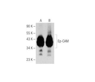
Anticorpo Ep-CAM (0.N.276): sc-71059
- Ep-CAM Antibody 0.N.276 è un monoclonale di topo IgG1 κ, citato in 7 pubblicazioni, fornito in 200 µg/ml
- testato contro la linea cellulare HG9 del carcinoma polmonare a piccole cellule di origine human
- raccomandato per il rilevamento di Ep-CAM di origine human in WB, IP, IF e FCM
- disponibile coniugato a phycoerythrin o a FITC per IF, IHC(P) e FCM
- Vedere Ep-CAM (C-10): sc-25308 for Ep-CAM per anticorpi coniugati, tra cui AC, HRP, FITC, PE, Alexa Fluor® 488, 594, 647, 680 e 790.
- m-IgG Fc BP-HRP e m-IgG1 BP-HRP sono i reagenti secondari preferiti per l'anticorpo Ep-CAM (0.N.276) for WB applications. Questi reagenti sono ora offerti in bundle con l'anticorpo Ep-CAM (0.N.276)(vedere le informazioni per l'ordine sotto).

LINK RAPIDI
VEDI ANCHE...
La molecola di adesione delle cellule epiteliali Ep-CAM, designata anche trasduttore del segnale di calcio associato al tumore 1 e MK-1, è una glicoproteina monomerica di membrana espressa nella maggior parte degli epiteli umani normali e nei carcinomi. Il gene Ep-CAM umano codifica una proteina di 314 aminoacidi che viene espressa in due forme, una maggiore e una minore, che si riducono con il trattamento con l'inibitore dell'amino-glicosilazione tunicamicina. L'Ep-CAM è sovraespressa in una varietà di carcinomi ed è quindi un potenziale bersaglio per la visualizzazione e la terapia dei tumori solidi umani. Ep-CAM contiene un dominio extracellulare contenente due ripetizioni simili al fattore di crescita epidermico, seguite da una regione povera di cisteina, necessaria per le proprietà di adesione della molecola.
Alexa Fluor® è un marchio registrato di Molecular Probes Inc., OR., USA
LI-COR® e Odyssey® sono marchi registrati di LI-COR Biosciences
Anticorpo Ep-CAM (0.N.276) Riferimenti:
- RNA aptamer contro un marcatore di cellule staminali tumorali, la molecola di adesione delle cellule epiteliali. | Shigdar, S., et al. 2011. Cancer Sci. 102: 991-8. PMID: 21281402
- Espressione, cristallizzazione e caratterizzazione preliminare ai raggi X dell'ectodominio della molecola di adesione cellulare epiteliale umana. | Pavšič, M. and Lenarčič, B. 2011. Acta Crystallogr Sect F Struct Biol Cryst Commun. 67: 1363-6. PMID: 22102232
- Demascheramento della molecola di adesione delle cellule epiteliali (EpCAM) sulle cellule tumorali epiteliali circolanti mediante trattamento con Tween®20 in pazienti con cancro al seno. | Hekimian, K., et al. 2012. Clin Chem Lab Med. 50: 701-8. PMID: 22505561
- Confronto tra mAbs che mirano alla molecola di adesione delle cellule epiteliali per il rilevamento delle metastasi linfonodali del cancro alla prostata con agenti di contrasto multimodali: PET/CT quantitativa su piccoli animali e NIRF. | Hall, MA., et al. 2012. J Nucl Med. 53: 1427-37. PMID: 22872743
- Espressione e ruolo della molecola di adesione delle cellule epiteliali nel nodulo displastico e nel carcinoma epatocellulare. | Bae, JS., et al. 2012. Int J Oncol. 41: 2150-8. PMID: 22993038
- Espressione della molecola di adesione delle cellule epiteliali e dell'antigene nucleare delle cellule proliferanti nell'epatocarcinogenesi indotta dalla dietilnitrosamina nei topi. | Kang, JS., et al. 2013. Exp Ther Med. 5: 138-142. PMID: 23251255
- La citocheratina 19, l'antigene carcinoembrionale e la molecola di adesione delle cellule epiteliali rilevano le metastasi linfonodali da cancro del polmone in campioni di aspirazione transbronchiale endobronchiale guidata da ultrasuoni. | Bugalho, A., et al. 2013. Clin Lung Cancer. 14: 704-12. PMID: 23886796
- La sovraespressione della molecola di adesione delle cellule epiteliali è associata a una prognosi più favorevole e alla risposta alla chemioterapia a base di platino nel carcinoma ovarico. | Woopen, H., et al. 2014. J Gynecol Oncol. 25: 221-8. PMID: 25045435
- Identificazione di microRNA specifici per le cellule tumorali positive alla molecola di adesione cellulare epiteliale nel carcinoma epatocellulare. | Ji, J., et al. 2015. Hepatology. 62: 829-40. PMID: 25953724
- Costruzione di un vettore di interferenza che ha come bersaglio il gene Ep-CAM e i suoi effetti sulla proliferazione delle cellule di cancro del colon-retto. | Qi, Y., et al. 2015. Drug Des Devel Ther. 9: 2647-52. PMID: 26028961
Informazioni ordini
| Nome del prodotto | Codice del prodotto | UNITÀ | Prezzo | Quantità | Preferiti | |
Ep-CAM Anticorpo (0.N.276) | sc-71059 | 200 µg/ml | $322.00 | |||
Ep-CAM (0.N.276): m-IgG Fc BP-HRP Bundle | sc-539340 | 200 µg Ab; 10 µg BP | $361.00 | |||
Ep-CAM (0.N.276): m-IgG1 BP-HRP Bundle | sc-541423 | 200 µg Ab; 20 µg BP | $361.00 | |||
Ep-CAM Anticorpo (0.N.276) FITC | sc-71059 FITC | 200 µg/ml | $336.00 | |||
Ep-CAM Anticorpo (0.N.276) PE | sc-71059 PE | 200 µg/ml | $349.00 |