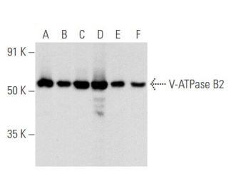
Date published: 2025-9-9

00800 4573 8000

SCBT Portrait Logo
Seach Input

Anticorps V-ATPase B2 (C-9): sc-166122

4.8(4)
Écrire une critiquePoser une question

Fiches techniques
  • L'Anticorps V-ATPase B2 (C-9) est un monoclonal de souris IgG3 κ, cité dans 4 publications, fourni en 200 µg/ml
  • spécifique d'un épitope situé entre les acides aminés 2-38 au niveau N-terminus de V-ATPase B2 d'origine human
  • recommandé pour la détection de V-ATPase B2 d'origine mouse, rat et human par WB, IP, IF et ELISA; également réactif avec d'autres espèces, dont et canine and bovine
  • m-IgG3 BP-HRP est le réactif de détection secondaire préféré pour V-ATPase B2 Anticorps (C-9) pour les applications WB. Ce réactif est désormais proposé dans un ensemble avec V-ATPase B2 Anticorps (C-9)(voir les informations de commande ci-dessous).

ACCÈS RAPIDE AUX LIENS

VOIR ÉGALEMENT...

Vacuolar-type H+-ATPase (V-ATPase) is a multisubunit enzyme responsible for acidification of eukaryotic intracellular organelles. V-ATPases pump protons against an electrochemical gradient, while F-ATPases reverse the process, thereby synthesizing ATP. A peripheral V1 domain, which is responsible for ATP hydrolysis, and a integral V0 domain, which is responsible for proton translocation, compose V-ATPase. Nine subunits (A-H) make up the V1 domain and five subunits (a, d, c, c' and c") make up the V0 domain. Like F-ATPase, V-ATPase most likely operates through a rotary mechanism. The V-ATPase V1 B subunit exists as two isoforms. In the inner ear, the V-ATPase B1 isoform functions in proton secretion and is required to maintain proper endolymph pH and normal auditory function. The gene encoding the human V-ATPase B1 isoform maps to chromosome 2cen-q13. Mutations in this gene cause distal renal tubular acidosis associated with sensorineural deafness. The V-ATPase B2 isoform is expressed in kidney and is the only B isoform expressed in osteoclasts. The gene encoding the human V-ATPase B2 isoform maps to chromosome 8p22-p21.

Pour la Recherche Uniquement. Non destiné à un usage diagnostique ou thérapeutique.

Alexa Fluor® est une marque déposée de Molecular Probes Inc., OR., USA

LI-COR® et Odyssey® sont marques déposées de LI-COR Biosciences

Anticorps V-ATPase B2 (C-9) Références:

  1. Distribution des Rab GTPases dans le rein de souris et comparaison avec la H+-ATPase vacuolaire.  |  Curtis, LM. and Gluck, S. 2005. Nephron Physiol. 100: p31-42. PMID: 15838183
  2. Relocalisation de la sous-unité B2 de la V-ATPase dans la membrane apicale des cellules claires de l'épididyme de souris déficientes en sous-unité B1.  |  Da Silva, N., et al. 2007. Am J Physiol Cell Physiol. 293: C199-210. PMID: 17392376
  3. Expression membranaire compensatoire de l'isoforme de la sous-unité B2 de la V-ATPase dans les cellules intercalaires médullaires rénales de souris déficientes en B1.  |  Paunescu, TG., et al. 2007. Am J Physiol Renal Physiol. 293: F1915-26. PMID: 17898041
  4. Rôle de CFTR et ClC-5 dans la modulation de l'activité de la H+-ATPase vacuolaire dans le tubule proximal du rein.  |  Carraro-Lacroix, LR., et al. 2010. Cell Physiol Biochem. 26: 563-76. PMID: 21063094
  5. La V-ATPase favorise la transition épithéliale-mésenchymateuse induite par le facteur de croissance transformant-β des cellules épithéliales tubulaires proximales de rat.  |  Cao, X., et al. 2012. Am J Physiol Renal Physiol. 302: F1121-32. PMID: 22129967
  6. Interaction entre la V-ATPase B2 et les récepteurs de la (Pro) rénine dans la promotion de la progression de la fibrose tubulo-interstitielle rénale.  |  Liu, Y., et al. 2016. Sci Rep. 6: 25035. PMID: 27121029
  7. L'altération de la V-ATPase entraîne une augmentation du pH lysosomal, une dégradation lysosomale perturbée et un blocage du flux autophagique, ce qui contribue à la neurotoxicité développementale induite par le fluorure.  |  Han, X., et al. 2022. Ecotoxicol Environ Saf. 236: 113500. PMID: 35421827
  8. La surexpression de la V-ATPase B2 atténue les lésions pulmonaires et la fibrose en stabilisant la perméabilisation de la membrane lysosomale et en augmentant la dégradation du collagène.  |  Lee, JU., et al. 2022. Exp Mol Med. 54: 662-672. PMID: 35624153
  9. La V-ATPase B2 favorise la phagocytose microgliale des débris de myéline en inactivant la voie de signalisation MAPK.  |  Li, Y., et al. 2024. Neuropeptides. 106: 102436. PMID: 38733728
  10. Le F-53B perturbe le métabolisme énergétique en inhibant l'axe V-ATPase-AMPK dans les cellules neuronales.  |  Zhang, Y., et al. 2025. J Hazard Mater. 487: 137111. PMID: 39793390

Informations pour la commande

Nom du produitRef. CatalogueCOND.Prix HTQTÉFavoris

Anticorps V-ATPase B2 (C-9)

sc-166122
200 µg/ml
$316.00

V-ATPase B2 (C-9): m-IgG3 BP-HRP Kit

sc-550390
200 µg Ab; 40 µg BP
$354.00

V-ATPase B2 (C-9) peptide neutralisant

sc-166122 P
100 µg/0.5 ml
$68.00

What application is the blocking peptide sc-166122 P appropriate for?

Posée par: cjMara
Thank you for your question. The blocking peptide is intended for use as a negative control, by pre-adsorbing the mouse monoclonal antibody against the antigen. For full protocol details, please contact our Technical Services Department or view our online protocol here: https://www.scbt.com/scbt/resources/protocols/peptide-neutralization
Répondue par: Technical Support
Date de publication: 2017-02-24
  • y_2025, m_8, d_27, h_5CST
  • bvseo_bulk, prod_bvqa, vn_bulk_3.0.42
  • cp_1, bvpage1
  • co_hasquestionsanswers, tq_1
  • loc_fr_FR, sid_166122, prod, sort_[SortEntry(order=LAST_APPROVED_ANSWER_SUBMISSION_TIME, direction=DESCENDING)]
  • clientName_scbt
  • bvseo_sdk, java_sdk, bvseo-4.0.0
  • CLOUD, getContent, 111ms
  • QUESTIONS, PRODUCT
Rated 5 de 5 de par Good Antibody for WBThis antibody is well-cited for a variety of applications including WB, IP, IF, and even ELISA. Strong, clear signal with little to no background. Works well in a variety of species including mouse, rat, and human samples.
Date de publication: 2017-01-27
Rated 5 de 5 de par Excellent immunofluorescence cytoplasmicExcellent immunofluorescence cytoplasmic and cytoplasmic vesicles staining in formalin-fixed HepG2 cells. -SCBT QC
Date de publication: 2015-05-18
Rated 5 de 5 de par Positive detection of endogenous VPositive detection of endogenous V-ATPase B2 expression in U-87 MG cells by WB. -SCBT QC
Date de publication: 2015-04-30
Rated 4 de 5 de par An increase in signal was observed via WesternAn increase in signal was observed via Western Blot when comparing mouse V-ATPase B2 transfected 293T cells with those that have not been transfected. -SCBT QC
Date de publication: 2015-03-11
  • y_2025, m_8, d_27, h_5
  • bvseo_bulk, prod_bvrr, vn_bulk_3.0.42
  • cp_1, bvpage1
  • co_hasreviews, tv_0, tr_4
  • loc_fr_FR, sid_166122, prod, sort_[SortEntry(order=SUBMISSION_TIME, direction=DESCENDING)]
  • clientName_scbt
  • bvseo_sdk, java_sdk, bvseo-4.0.0
  • CLOUD, getReviews, 14ms
  • REVIEWS, PRODUCT
Anticorps V-ATPase B2 (C-9) est évalué 4.8 de 5 de 4.
  • y_2025, m_8, d_27, h_5
  • bvseo_bulk, prod_bvrr, vn_bulk_3.0.42
  • cp_1, bvpage1
  • co_hasreviews, tv_0, tr_4
  • loc_fr_FR, sid_166122, prod, sort_[SortEntry(order=SUBMISSION_TIME, direction=DESCENDING)]
  • clientName_scbt
  • bvseo_sdk, java_sdk, bvseo-4.0.0
  • CLOUD, getAggregateRating, 103ms
  • REVIEWS, PRODUCT