")
SCF Anticorpo (G-3): sc-13126
- SCF Anticorpo G-3é um anticorpo monoclonal produzido em camundongo IgG2b κ SCF anticorpo, citado em 53 publicações, fornecido em 200 µg/ml
- Produzido contra aminoácidos 26-214 de stem cell factor (SCF) de human origem
- SCF Anticorpo (G-3) é recomendado para a detecção de SCF de mouse, rat e human origem em WB, IP, IF, IHC(P) e ELISA
- Anticorpo anti-SCF O anticorpo anti-SCF (G-3) está disponível conjugado com agarose para IP; HRP para WB, IHC(P) e ELISA; e com fioeritrina ou FITC para IF, IHC(P) e FCM
- também disponível conjugado com Alexa Fluor® 488, Alexa Fluor® 546, Alexa Fluor® 594 ou Alexa Fluor® 647 para WB (RGB), IF, IHC(P) e FCM, e para utilização com sistemas de imagiologia fluorescente RGB, tais como iBright™ FL1000, FluorChem™, Typhoon, Azure e outros sistemas comparáveis
- também disponível conjugado com Alexa Fluor® 680 ou Alexa Fluor® 790 para WB (NIR), IF e FCM; para utilização com sistemas de deteção de infravermelhos próximos (NIR), tais como LI-COR®Odyssey®, iBright™ FL1000, FluorChem™, Typhoon, Azure e outros sistemas comparáveis
- 2b BP-HRP">m-IgG2b BP-HRP e m-IgGκ BP-HRP são os reagentes de deteção secundários preferidos para SCF Antibody (G-3) para aplicações WB e IHC(P). Estes reagentes são agora oferecidos em conjuntos com SCF Antibody (G-3)(ver informações de encomenda abaixo).

LINKS RÁPIDOS
VEJA TAMBÉM
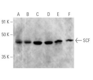
O Anticorpo SCF (G-3) é um anticorpo monoclonal de cadeia leve IgG2b kappa de camundongo que detecta a proteína SCF de origem de camundongo, rato e humana por WB, IP, IF, IHCP e ELISA. O anticorpo SCF (G-3) está disponível em formas não conjugadas e em várias formas conjugadas, incluindo agarose, HRP, PE, FITC e vários conjugados Alexa Fluor®. O SCF desempenha um papel crucial como fixador do recetor transmembranar da tirosina quinase proto-oncogene c-Kit, que é vital para vários processos celulares. O SCF é uma citocina pleiotrópica que existe em duas formas alternativas emendadas, consistindo em 248 e 220 aminoácidos nos sistemas humano e do camundongo, respetivamente. A forma maior de SCF é expressa predominantemente em fibroblastos, no cérebro e no timo, enquanto a variante mais pequena se encontra em tecidos como o baço, o testículo, a placenta e o cerebelo. O SCF é essencial para o desenvolvimento de células germinativas, células progenitoras hematopoiéticas e precursores de melanócitos, uma vez que o SCF estimula a proliferação e a maturação de mastócitos maduros e imaturos. A interação do SCF com o c-Kit é crítica para vários processos biológicos, incluindo a hematopoiese e a pigmentação, tornando o anticorpo monoclonal SCF (G-3) uma ferramenta valiosa para a investigação em biologia do desenvolvimento e estudos do cancro.
Alexa Fluor® é uma marca comercial da Molecular Probes Inc., OR., EUA
LI-COR® e Odyssey® são marcas registadas da LI-COR Biosciences
Referencias do SCF Anticorpo (G-3):
- Expressão e processamento diferenciais de duas formas associadas a células do ligando kit: KL-1 e KL-2. | Huang, EJ., et al. 1992. Mol Biol Cell. 3: 349-62. PMID: 1378327
- Apoio à hematopoiese humana em culturas de medula óssea a longo prazo por células estromais murinas que expressam seletivamente as formas ligadas à membrana e segregadas do homólogo humano do produto do gene steel, o fator de células estaminais. | Toksoz, D., et al. 1992. Proc Natl Acad Sci U S A. 89: 7350-4. PMID: 1380155
- A clivagem de factores de crescimento ancorados na membrana envolve actividades de proteases distintas reguladas por mecanismos comuns. | Pandiella, A., et al. 1992. J Biol Chem. 267: 24028-33. PMID: 1385433
- O fator de crescimento dos mastócitos está localizado perto do locus steel no cromossoma 10 do rato e é eliminado em vários alelos steel. | Copeland, NG., et al. 1990. Cell. 63: 175-83. PMID: 1698554
- Estrutura primária e expressão funcional de DNAs de factores de células estaminais de rato e humanas. | Martin, FH., et al. 1990. Cell. 63: 203-11. PMID: 2208279
- Papel do fator de células estaminais no nicho placentário. | Khodadi, E., et al. 2016. Cell Tissue Res. 366: 523-531. PMID: 27234501
- Eritropoietina, fator de células estaminais e migração de células cancerosas. | Vazquez-Mellado, MJ., et al. 2017. Vitam Horm. 105: 273-296. PMID: 28629522
- Os adipócitos da medula óssea promovem a regeneração das células estaminais e a hematopoiese através da secreção de SCF. | Zhou, BO., et al. 2017. Nat Cell Biol. 19: 891-903. PMID: 28714970
- Fator de células estaminais: a ponte entre os adipócitos da medula óssea e as células hematopoiéticas. | Li, Z. and MacDougald, OA. 2019. Haematologica. 104: 1689-1691. PMID: 31473604
- A inibição do fator solúvel das células estaminais promove a reparação da mucosa intestinal. | Garcia-Hernandez, V., et al. 2023. Inflamm Bowel Dis. 29: 1133-1144. PMID: 36688460
- O fator de células estaminais e o cKIT modulam a glicólise endotelial em hipoxia. | Jeong, H., et al. 2024. Cardiovasc Res. 120: 745-755. PMID: 38507654
- Os monócitos não produzem mastócitos quando cultivados na presença de SCF. Caracterização do progenitor de mastócitos circulante como uma célula formadora de colónias c-kit+, CD34+, Ly-, CD14-, CD17-. | Agis, H., et al. 1993. J Immunol. 151: 4221-7. PMID: 7691941
Informacoes sobre ordens
| Nome do Produto | Numero de Catalogo | UNID | Preco | Qde | FAVORITOS | |
SCF Anticorpo (G-3) | sc-13126 | 200 µg/ml | $322.00 | |||
Pacote do SCF (G-3): m-IgGκ BP-HRP | sc-520598 | 200 µg Ab, 40 µg BP | $361.00 | |||
Pacote do SCF (G-3): m-IgG2b BP-HRP | sc-548841 | 200 µg Ab; 10 µg BP | $361.00 | |||
SCF Anticorpo (G-3) AC | sc-13126 AC | 500 µg/ml, 25% agarose | $424.00 | |||
SCF Anticorpo (G-3) HRP | sc-13126 HRP | 200 µg/ml | $322.00 | |||
SCF Anticorpo (G-3) FITC | sc-13126 FITC | 200 µg/ml | $336.00 | |||
SCF Anticorpo (G-3) PE | sc-13126 PE | 200 µg/ml | $349.00 | |||
SCF Anticorpo (G-3) Alexa Fluor® 488 | sc-13126 AF488 | 200 µg/ml | $364.00 | |||
SCF Anticorpo (G-3) Alexa Fluor® 546 | sc-13126 AF546 | 200 µg/ml | $364.00 | |||
SCF Anticorpo (G-3) Alexa Fluor® 594 | sc-13126 AF594 | 200 µg/ml | $364.00 | |||
SCF Anticorpo (G-3) Alexa Fluor® 647 | sc-13126 AF647 | 200 µg/ml | $364.00 | |||
SCF Anticorpo (G-3) Alexa Fluor® 680 | sc-13126 AF680 | 200 µg/ml | $364.00 | |||
SCF Anticorpo (G-3) Alexa Fluor® 790 | sc-13126 AF790 | 200 µg/ml | $364.00 |