")
MAVS抗体(E-3): sc-166583
- MAVS抗体 E-3はマウスモノクローナルIgG2aMAVS 抗体 です。200 µg/mlで提供
- アミノ酸に対して1-135マッピングされたN-terminal細胞質ドメイン内のMAVSのhuman起源
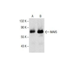
- MAVS抗体 (E-3) human由来のMAVS WB, IP, IF, IHC(P) と ELISAでの検出にはお勧めします
- 抗 MAVS 抗体 (E-3) は、IP 用には アガロース、WB、IHC(P)、ELISA 用には HRP、IF、IHC(P)、FCM 用には フィコエリスリン または FITC にそれぞれ結合したものが利用可能
- WB (RGB)、IF、IHC(P)、FCM、iBright™ FL1000、FluorChem™、Typhoon、Azureと他の同等システムでRGB蛍光イメージングシステム用のAlexa Fluor® 488、Alexa Fluor® 546、Alexa Fluor® 594 または Alexa Fluor® 647、に共役での利用可能です。
- WB (NIR)、IF、FCMとLI-COR®/Odyssey®、iBright™ FL1000、FluorChem™、Typhoon、Azureと他の同等システムで近赤外(NIR)検出法用のAlexa Fluor® 680 または Alexa Fluor® 790、に共役での利用可能です。
- m-IgG Fc BP-HRP、 2a BP-HRP">m-IgG2a BP-HRPおよびm-IgGκ BP-HRPは、MAVS Antibody (E-3) WBおよびIHC(P)アプリケーション用。 の二次検出試薬として推奨されています。これらの試薬は現在、MAVS Antibody (E-3) とバンドルして提供されています(下記の注文情報を参照)。

クイックリンク
関連項目
MAVS 抗体 (E-3) は IgG2aκ マウスモノクローナル MAVS 抗体(IPS1 抗体、VISA 抗体とも呼ばれる)で、ヒト由来の MAVS タンパク質を WB、IP、IF、IHC(P)、ELISA で検出します。MAVS抗体(E-3)は、ノンコンジュゲートの抗MAVS抗体の他、アガロース、HRP、PE、FITC、複数のAlexa Fluor®コンジュゲートなど、様々なコンジュゲートタイプの抗MAVS抗体としてご利用いただけます。MAVS (mitochondrial antiviral-signaling protein) は、IPS1、KIAA1271、VISA、CARDIF としても知られ、1つのCARDドメインと複数の膜貫通ドメインを含む540アミノ酸のタンパク質で、ミトコンドリア外膜に局在する。全身に発現し、肝臓、心臓、胎盤、骨格筋、末梢血白血球で最も発現が高い。MAVSは、二本鎖(ds)ウイルス複製を検出するRIG-Iなどのタンパク質の下流で機能し、dsウイルス感染に対する適切な免疫再反応に必要である。MAVSは、抗ウイルス性サイトカインの誘導につながる経路を活性化し、ウイルスが誘導するアポトーシスから細胞を保護すると考えられている。MAVSの機能は、CARDドメインと膜貫通ドメインを分解するプロテアーゼ複合体による切断によって不活性化され、それによってMAVSが他のタンパク質と相互作用するのを防ぐことができる。MAVSは代替スプライシングにより3つのアイソフォームが発現している。
Alexa Fluor® はMolecular Probes Inc., OR., USAの商標です。
LI-COR® and Odyssey® はLI-COR Biosciencesの登録商標です。
MAVS抗体(E-3) 参考文献:
- C型肝炎ウイルスのプロテアーゼNS3/4Aは, ミトコンドリアの抗ウイルスシグナル伝達タンパク質を切断し, 自然免疫を回避する。 | Li, XD., et al. 2005. Proc Natl Acad Sci U S A. 102: 17717-22. PMID: 16301520
- C型肝炎ウイルス感染におけるIFN-βプロモーター刺激因子1のウイルス制御と治療制御。 | Loo, YM., et al. 2006. Proc Natl Acad Sci U S A. 103: 6001-6. PMID: 16585524
- NS3プロテアーゼ依存性および非依存性メカニズムによるC型肝炎ウイルス感染細胞におけるdsRNA誘導シグナル伝達の阻害。 | Cheng, G., et al. 2006. Proc Natl Acad Sci U S A. 103: 8499-504. PMID: 16707574
- C型肝炎ウイルスNS3-4Aのタンパク質分解によるMAVS/IPS-1/VISA/Cardif-IKKepsilon分子複合体のミトコンドリア外膜からの解離。 | Lin, R., et al. 2006. J Virol. 80: 6072-83. PMID: 16731946
- TRAF3とCardifの直接的かつ特異的な相互作用による抗ウイルス応答の制御。 | Saha, SK., et al. 2006. EMBO J. 25: 3257-63. PMID: 16858409
- レジオネラ・ニューモフィラは, IPS-1とIRF3を介して肺上皮細胞にIFNβを誘導し, 細菌の複製も制御している。 | Opitz, B., et al. 2006. J Biol Chem. 281: 36173-9. PMID: 16984921
- GBウイルスBは, NS3/4Aが介在するアダプタータンパク質MAVSの切断により, RIG-Iシグナル伝達を阻害する。 | Chen, Z., et al. 2007. J Virol. 81: 964-76. PMID: 17093192
- 腸管上皮細胞におけるRIG-I/IPS-1を介したシグナル伝達による自然免疫防御機構の活性化。 | Hirata, Y., et al. 2007. J Immunol. 179: 5425-32. PMID: 17911629
- RIGI/MAVS経路シグナル伝達を阻害するMAVSスプライシングバリアントの同定。 | Lad, SP., et al. 2008. Mol Immunol. 45: 2277-87. PMID: 18207245
- 乳酸はMAVSを標的とするRLRシグナル伝達の自然な抑制因子である。 | Zhang, W., et al. 2019. Cell. 178: 176-189.e15. PMID: 31155231
- MAVSはグルコース代謝とRIG-I様受容体シグナル伝達を統合する。 | He, QQ., et al. 2023. Nat Commun. 14: 5343. PMID: 37660168
- エピジェネティックな撹乱後のCPT1A誘導は、MAVSのパルミトイル化と活性化を促進し、抗腫瘍免疫を増強する。 | Zhang, G., et al. 2023. Mol Cell. 83: 4370-4385.e9. PMID: 38016475
注文情報
| 製品名 | カタログ # | 単位 | 価格 | 数量 | お気に入り | |
MAVS 抗体 (E-3) | sc-166583 | 200 µg/ml | $322.00 | |||
MAVS (E-3): m-IgG Fc BP-HRP Bundle | sc-528998 | 200 µg Ab; 10 µg BP | $361.00 | |||
MAVS (E-3): m-IgGκ BP-HRP Bundle | sc-521667 | 200 µg Ab, 40 µg BP | $361.00 | |||
MAVS (E-3): m-IgG2a BP-HRP Bundle | sc-547231 | 200 µg Ab; 10 µg BP | $361.00 | |||
MAVS 抗体 (E-3) AC | sc-166583 AC | 500 µg/ml, 25% agarose | $424.00 | |||
MAVS 抗体 (E-3) HRP | sc-166583 HRP | 200 µg/ml | $322.00 | |||
MAVS 抗体 (E-3) FITC | sc-166583 FITC | 200 µg/ml | $336.00 | |||
MAVS 抗体 (E-3) PE | sc-166583 PE | 200 µg/ml | $349.00 | |||
MAVS 抗体 (E-3) Alexa Fluor® 488 | sc-166583 AF488 | 200 µg/ml | $364.00 | |||
MAVS 抗体 (E-3) Alexa Fluor® 546 | sc-166583 AF546 | 200 µg/ml | $364.00 | |||
MAVS 抗体 (E-3) Alexa Fluor® 594 | sc-166583 AF594 | 200 µg/ml | $364.00 | |||
MAVS 抗体 (E-3) Alexa Fluor® 647 | sc-166583 AF647 | 200 µg/ml | $364.00 | |||
MAVS 抗体 (E-3) Alexa Fluor® 680 | sc-166583 AF680 | 200 µg/ml | $364.00 | |||
MAVS 抗体 (E-3) Alexa Fluor® 790 | sc-166583 AF790 | 200 µg/ml | $364.00 |