")
-D-galactose, CAS Number: 7368-73-2")
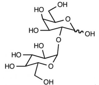
2-O-(α-D-Glucopyranosyl)-D-galactose (CAS 7368-73-2)
QUICK LINKS
2-O-(α-D-Glucopyranosyl)-D-galactose is a key molecule in glycobiology research, primarily investigated for its role in carbohydrate metabolism and cellular recognition processes. This chemical acts as a glycosyl donor or acceptor in enzymatic reactions mediated by glycosyltransferases, contributing to the biosynthesis of complex oligosaccharides and glycoconjugates. In research, it has been utilized as a substrate in enzymatic assays to characterize the specificity and kinetics of glycosyltransferase enzymes involved in the biosynthesis of glycan structures. Additionally, 2-O-(α-D-Glucopyranosyl)-D-galactose has been studied for its involvement in cell-cell interactions and molecular recognition events mediated by glycan-binding proteins, such as lectins. Its presence on cell surfaces or as part of extracellular glycoconjugates may play a role in various biological processes, including cell adhesion, immune response modulation, and pathogen recognition. Furthermore, researchers have investigated the structural features and biosynthetic pathways associated with 2-O-(α-D-Glucopyranosyl)-D-galactose-containing glycoconjugates to understand their physiological significance in cellular communication and disease processes. Overall, this chemical serves as a valuable tool for studying glycan biosynthesis, glycan-mediated interactions, and their implications in diverse biological contexts.
2-O-(α-D-Glucopyranosyl)-D-galactose (CAS 7368-73-2) References
- Substrate recognition by the collagen-binding domain of Clostridium histolyticum class I collagenase. | Matsushita, O., et al. 2001. J Biol Chem. 276: 8761-70. PMID: 11121400
- Crystal structure and substrate-binding mode of GH63 mannosylglycerate hydrolase from Thermus thermophilus HB8. | Miyazaki, T., et al. 2015. J Struct Biol. 190: 21-30. PMID: 25712767
- Crystal structure of the enzyme-product complex reveals sugar ring distortion during catalysis by family 63 inverting α-glycosidase. | Miyazaki, T., et al. 2016. J Struct Biol. 196: 479-486. PMID: 27688023
- Incorporation of 'click' chemistry glycomimetics dramatically alters triple-helix stability in an adiponectin model peptide. | Lutteroth, KR., et al. 2017. Org Biomol Chem. 15: 5602-5608. PMID: 28639641
- Chemical Synthesis and Biological Evaluations of Adiponectin Collagenous Domain Glycoforms. | Wu, H., et al. 2021. J Am Chem Soc. 143: 7808-7818. PMID: 33979146
- Recent advances in chemical synthesis of O-linked glycopeptides and glycoproteins: An advanced synthetic tool for exploring the biological realm. | Zhao, J., et al. 2023. Curr Opin Chem Biol. 77: 102405. PMID: 37897925
- The structure of the disaccharide unit of the renal glomerular basement membrane. | Spiro, RG. 1967. J Biol Chem. 242: 4813-23. PMID: 6070267
- Isolation of a collagen fraction from the body-wall glycoproteins of the leech (Hirudo medicinalis), and characterization of its carbohydrate--amino acid portion. | Biswas, T. and Mukherjee, AK. 1978. Carbohydr Res. 63: 173-81. PMID: 667877
- The o-acetyl groups of the specific substance from Pneumococcus type 34 (U.S. type 41)☆ | J.R. Dixon 1, W.K. Roberts 2, G.T. Mills 3, J.G. Buchanan, J. Baddiley. 1968. Carbohydrate Research. 8: 262-265.
Ordering Information
| Product Name | Catalog # | UNIT | Price | Qty | FAVORITES | |
2-O-(α-D-Glucopyranosyl)-D-galactose, 5 mg | sc-220754 | 5 mg | $650.00 |