")
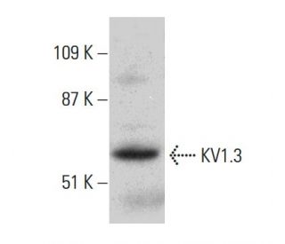
: sc-398855. Analisi Western blot dell'espressione di KV1.3 nel lisato di cellule intere Jurkat.")
: sc-398855. Analisi in Western blot dell'espressione di KV1.3 in lisati di cellule intere non trasfettate 293T: sc-117752 (A), trasfettate con KV1.3 umano 293T: sc-159570 (B) e Jurkat (C).")
Anticorpo KV1.3 (G-9): sc-398855
- KV1.3 Antibody G-9 è un monoclonale di topo IgG3 KV1.3 antibody, citato in 2 pubblicazioni, fornito in 200 µg/ml
- specifico per un epitopo mapping tra gli aminoacidi 535-556 all´interno di un dominio citoplasmatico C-terminal dell´KV1.3 di origine human
- raccomandato per il rilevamento di KV1.3 di origine mouse, rat e human in WB, IP, IF e ELISA
- m-IgG Fc BP-HRP è il reagente secondario di rilevazione preferito per l'anticorpo KV1.3 (G-9) per applicazioni WB. Questo reagente è ora offerto in combinazione con l'anticorpo KV1.3 (G-9)(vedere le informazioni per l'ordine qui sotto).

LINK RAPIDI
VEDI ANCHE...
L'anticorpo KV1.3 (G-9) è un anticorpo monoclonale IgG3 di topo contro la proteina KV1.3 (designata anche come anticorpo KV1.3) che rileva la proteina KV1.3 di origine murina, ratta e umana mediante WB, IP, IF ed ELISA. L'anticorpo KV1.3 (G-9) è disponibile come anticorpo anti-KV1.3 non coniugato. I canali K+ voltaggio-gati nella membrana plasmatica controllano la ripolarizzazione e la frequenza dei potenziali d'azione nei neuroni, nei muscoli e in altre cellule eccitabili. La famiglia dei geni KV codifica più di 30 geni che comprendono le subunità dei canali K+ e variano nelle loro proprietà di gating e permeazione, nella distribuzione subcellulare e nei modelli di espressione. I canali KV funzionali si assemblano come tetrameri costituiti da subunità α formanti il poro (KVα), che includono le proteine KV1, KV2, KV3 e KV4, e da subunità accessorie o KVβ che modificano le proprietà di gating delle subunità KVα coespresse. Esistono differenze nei modelli di traffico, elaborazione biosintetica ed espressione di superficie delle principali subunità KV1 (KV1.1, KV1.2, KV1.4, KV1.5 e KV1.6) espresse nel cervello del ratto e dell'uomo, suggerendo che le singole subunità proteiche sono altamente regolate per controllare l'assemblaggio e la formazione di canali neuronali funzionali.
Alexa Fluor® è un marchio registrato di Molecular Probes Inc., OR., USA
LI-COR® e Odyssey® sono marchi registrati di LI-COR Biosciences
Anticorpo KV1.3 (G-9) Riferimenti:
- Composizione di subunità dei canali Kv1 nel SNC umano. | Coleman, SK., et al. 1999. J Neurochem. 73: 849-58. PMID: 10428084
- La composizione delle subunità determina l'espressione della superficie del canale del potassio Kv1. | Manganas, LN. and Trimmer, JS. 2000. J Biol Chem. 275: 29685-93. PMID: 10896669
- I canali Kv1.3 facilitano la connessione tra metabolismo e flusso sanguigno nel cuore. | Ohanyan, V., et al. 2017. Microcirculation. 24: PMID: 28504408
- Kv1.3 modula la neuroinfiammazione e la neurodegenerazione nella malattia di Parkinson. | Sarkar, S., et al. 2020. J Clin Invest. 130: 4195-4212. PMID: 32597830
- Fisiologia del canale K+ Kv1.3 valutata mediante modulazione genetica e farmacologica. | Varanita, T., et al. 2023. Physiology (Bethesda). 38: 0. PMID: 35998249
- Il blocco del canale del potassio Kv1.3 inibisce la neuroinfiammazione mediata dalla microglia nell'epilessia. | Zhang, X., et al. 2022. Int J Mol Sci. 23: PMID: 36499018
- Kv1.3 sotto i riflettori per il trattamento delle malattie immunitarie. | Navarro-Pérez, M., et al. 2024. Expert Opin Ther Targets. 28: 67-82. PMID: 38316438
- Modulazione farmacologica di Kv1.3 dipendente da KCNE4. | Sastre, D., et al. 2024. Biochem Pharmacol. 226: 116368. PMID: 38880360
- Il canale del potassio Kv1.1 del cervello: studi in vitro e in vivo sull'assemblaggio delle subunità e sull'elaborazione post-traslazionale. | Deal, KK., et al. 1994. J Neurosci. 14: 1666-76. PMID: 8126562
- Localizzazione immunoistochimica di cinque membri delle subunità del canale Kv1: localizzazione subcellulare contrastante e co-localizzazione neurone-specifica nel cervello di ratto. | Veh, RW., et al. 1995. Eur J Neurosci. 7: 2189-205. PMID: 8563969
- Le subunità beta promuovono l'espressione della superficie dei canali K+ attraverso effetti precoci nella biosintesi. | Shi, G., et al. 1996. Neuron. 16: 843-52. PMID: 8608002
- Associazione e colocalizzazione delle subunità beta Kvbeta1 e Kvbeta2 con le subunità alfa Kv1 nei complessi dei canali K+ del cervello di mammifero. | Rhodes, KJ., et al. 1997. J Neurosci. 17: 8246-58. PMID: 9334400
Informazioni ordini
| Nome del prodotto | Codice del prodotto | UNITÀ | Prezzo | Quantità | Preferiti | |
KV1.3 Anticorpo (G-9) | sc-398855 | 200 µg/ml | $322.00 | |||
KV1.3 (G-9): m-IgG Fc BP-HRP Bundle | sc-526332 | 200 µg Ab; 10 µg BP | $361.00 | |||
KV1.3 (G-9) peptide neutralizzante | sc-398855 P | 100 µg/0.5 ml | $69.00 |