")
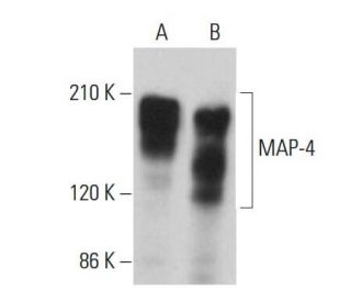
MAP-4 Antibody (F-3): sc-365187
- MAP-4 Antibody (F-3) is a mouse monoclonal IgG1 κ provided at 200 µg/ml
- raised against amino acids 1-300 mapping at the N-terminus of MAP-4 of human origin
- recommended for detection of MAP-4 isoforms 1 and 2 of human origin by WB, IP, IF and ELISA
- m-IgG Fc BP-HRP, m-IgG1 BP-HRP and m-IgGκ BP-HRP are the preferred secondary detection reagents for MAP-4 Antibody (F-3) for WB applications. These reagents are now offered in bundles with MAP-4 Antibody (F-3) (see ordering information below).

MAP-4 Antibody (F-3) is a mouse monoclonal IgG1 antibody that detects MAP-4 in human samples through western blotting (WB), immunoprecipitation (IP), immunofluorescence (IF), and enzyme-linked immunosorbent assay (ELISA). MAP-4 is a crucial microtubule-associated protein that plays a significant role in the assembly and stability of microtubules, which are essential components of the cytoskeletal network. MAP-4 is primarily expressed in vital organs such as the kidney, lung, liver, testis, and spleen, where MAP-4 contributes to the organization and dynamics of the microtubule system. Notably, MAP-4 contains three 18-amino acid repeats that are homologous to those found in other microtubule-associated proteins, highlighting MAP-4′s structural importance in facilitating microtubule assembly and influencing the spatial distribution of microtubules within cells. Proper functioning of MAP-4 is critical for various cellular processes, including the formation of the mitotic spindle during cell division, which ensures accurate chromosome segregation. Disruptions in MAP-4 function can lead to cellular instability and have been implicated in various diseases, making MAP-4 a vital target for research in cell biology and pathology. Anti-MAP-4 antibody (F-3) is an invaluable tool for researchers aiming to explore the intricate roles of microtubule dynamics in health and disease.
Alexa Fluor® is a trademark of Molecular Probes Inc., OR., USA
LI-COR® and Odyssey® are registered trademarks of LI-COR Biosciences
MAP-4 Antibody (F-3) References:
- Microtubule-associated protein 4 (MAP4) regulates assembly, protomer-polymer partitioning and synthesis of tubulin in cultured cells. | Nguyen, HL., et al. 1999. J Cell Sci. 112 (Pt 12): 1813-24. PMID: 10341201
- Truncation of the projection domain of MAP4 (microtubule-associated protein 4) leads to attenuation of microtubule dynamic instability. | Permana, S., et al. 2005. Cell Struct Funct. 29: 147-57. PMID: 15840946
- An isoform of microtubule-associated protein 4 inhibits kinesin-driven microtubule gliding. | Tokuraku, K., et al. 2007. J Biochem. 141: 585-91. PMID: 17317690
- Distinct neuronal localization of microtubule-associated protein 4 in the mammalian brain. | Tokuraku, K., et al. 2010. Neurosci Lett. 484: 143-7. PMID: 20727942
- The p38/mitogen-activated protein kinase pathway is implicated in lipopolysaccharide-induced microtubule depolymerization via up-regulation of microtubule-associated protein 4 phosphorylation in human vascular endothelium. | Zhou, Z., et al. 2015. Surgery. 157: 590-8. PMID: 25633728
- Cardiac proteomics reveals the potential mechanism of microtubule associated protein 4 phosphorylation-induced mitochondrial dysfunction. | Li, L., et al. 2019. Burns Trauma. 7: 8. PMID: 30906793
- Cyclin B interaction with microtubule-associated protein 4 (MAP4) targets p34cdc2 kinase to microtubules and is a potential regulator of M-phase microtubule dynamics. | Ookata, K., et al. 1995. J Cell Biol. 128: 849-62. PMID: 7876309
- Cellular microtubules heterogeneous in their content of microtubule-associated protein 4 (MAP4). | Chapin, SJ. and Bulinski, JC. 1994. Cell Motil Cytoskeleton. 27: 133-49. PMID: 7909279
- A muscle-specific variant of microtubule-associated protein 4 (MAP4) is required in myogenesis. | Mangan, ME. and Olmsted, JB. 1996. Development. 122: 771-81. PMID: 8631255
- Phosphorylation of microtubule-associated proteins MAP2 and MAP4 by the protein kinase p110mark. Phosphorylation sites and regulation of microtubule dynamics. | Illenberger, S., et al. 1996. J Biol Chem. 271: 10834-43. PMID: 8631898
Ordering Information
| Product Name | Catalog # | UNIT | Price | Qty | FAVORITES | |
MAP-4 Antibody (F-3) | sc-365187 | 200 µg/ml | $322.00 | |||
MAP-4 Antibody (F-3): m-IgG Fc BP-HRP Bundle | sc-537786 | 200 µg Ab; 10 µg BP | $361.00 | |||
MAP-4 Antibody (F-3): m-IgGκ BP-HRP Bundle | sc-535172 | 200 µg Ab; 40 µg BP | $361.00 | |||
MAP-4 Antibody (F-3): m-IgG1 BP-HRP Bundle | sc-545351 | 200 µg Ab; 20 µg BP | $361.00 |