")
</a> que nous recommandons (données d'échantillonnage indiquées).")
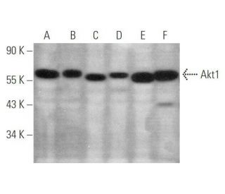
Cet anticorps polyclonal n'est plus commercialisé. Voir l'anticorps monoclonal Akt1 (B-1) que nous recommandons (données d'échantillonnage indiquées).
Anticorps Akt1 (C-20): sc-1618
- L'anticorps Akt1 C-20 est un polyclonal IgG de chèvre fourni en 200 µg/ml; ou IgG de lapin; 200 µg/ml, sc-1618-R
- épitope situé au niveau C-terminus de Akt1 d'origine human
- Anticorps polyclonal arrêté

Pour la Recherche Uniquement. Non destiné à un usage diagnostique ou thérapeutique.
Alexa Fluor® est une marque déposée de Molecular Probes Inc., OR., USA
Informations pour la commande
Akt1 (B-1): sc-5298 [ Monoclonal recommandé en remplacement de Akt1 (C-20) ]
| Nom du produit | Ref. Catalogue | COND. | Prix HT | QTÉ | Favoris | |
Anticorps Akt1 (B-1) | sc-5298 | 200 µg/ml | $322.00 | |||
Akt1 (B-1): m-IgG Fc BP-HRP Kit | sc-528153 | 200 µg Ab; 10 µg BP | $361.00 | |||
Akt1 (B-1): m-IgGκ BP-HRP Kit | sc-520461 | 200 µg Ab, 40 µg BP | $361.00 | |||
Akt1 (B-1): m-IgG1 BP-HRP Kit | sc-542789 | 200 µg Ab; 20 µg BP | $361.00 | |||
Anticorps Akt1 (B-1) AC | sc-5298 AC | 500 µg/ml, 25% agarose | $424.00 | |||
Anticorps Akt1 (B-1) HRP | sc-5298 HRP | 200 µg/ml | $322.00 | |||
Anticorps Akt1 (B-1) FITC | sc-5298 FITC | 200 µg/ml | $336.00 | |||
Anticorps Akt1 (B-1) PE | sc-5298 PE | 200 µg/ml | $349.00 | |||
Anticorps Akt1 (B-1) Alexa Fluor® 488 | sc-5298 AF488 | 200 µg/ml | $364.00 | |||
Anticorps Akt1 (B-1) Alexa Fluor® 546 | sc-5298 AF546 | 200 µg/ml | $364.00 | |||
Anticorps Akt1 (B-1) Alexa Fluor® 594 | sc-5298 AF594 | 200 µg/ml | $364.00 | |||
Anticorps Akt1 (B-1) Alexa Fluor® 647 | sc-5298 AF647 | 200 µg/ml | $364.00 | |||
Anticorps Akt1 (B-1) Alexa Fluor® 680 | sc-5298 AF680 | 200 µg/ml | $364.00 | |||
Anticorps Akt1 (B-1) Alexa Fluor® 790 | sc-5298 AF790 | 200 µg/ml | $364.00 |