")
</a> monoclonal antibody (sample data shown).")
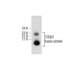
This polyclonal antibody has been discontinued. See our recommended TGFβ2 (H-6) monoclonal antibody (sample data shown).
TGFβ2 Antibody (V): sc-90
- TGFβ2 Antibody (V) is a rabbit polyclonal IgG; 200 µg/ml
- epitope mapping at the C-terminus of TGFβ2 of human origin
- Discontinued polyclonal antibody

QUICK LINKS
Description
For Research Use Only. Not Intended for Diagnostic or Therapeutic Use.
Alexa Fluor® is a trademark of Molecular Probes Inc., OR., USA
Ordering Information
TGFβ2 (H-6): sc-374659 [ Recommended monoclonal replacement for TGFβ2 (V) ]
| Product Name | Catalog # | UNIT | Price | Qty | FAVORITES | |
TGFβ2 Antibody (H-6) | sc-374659 | 200 µg/ml | $322.00 | |||
TGFβ2 Antibody (H-6): m-IgG3 BP-HRP Bundle | sc-550583 | 200 µg Ab; 40 µg BP | $361.00 | |||
TGFβ2 (H-6) Neutralizing Peptide | sc-374659 P | 100 µg/0.5 ml | $69.00 |