")
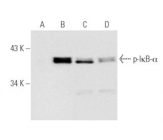
 et induites par le TNF-α (B,D). Les anticorps testés sont l'anticorps p-IκB-α (B-9): sc-8404 (A,B) et l'anticorps IκB-α (H-4): sc-1643 (C,D).")
: sc-8404. Coloration par immunofluorescence de cellules HeLa fixées au méthanol et traitées au TNFa, montrant la localisation nucléaire de l'IkB-a activé.")
: sc-8404. Coloration par immunoperoxydase d'un carcinome mammaire humain fixé au formol et inclus en paraffine, montrant la localisation nucléaire de l'IkB-a activé.")
Anticorps p-NFKBIA/IkB alpha (B-9): sc-8404
- L'Anticorps p-NFKBIA/IkB alpha (B-9) est un monoclonal de souris IgG2b κ, cité dans 524 publications, fourni en 200 µg/ml
- Anti-L'Anticorps p-NFKBIA/IkB alpha (B-9) est recommandé pour la détection de IκB-α phosphorylated at Ser 32 d'origine mouse, rat et human par WB, IP, IF et IHC(P)
- Anti-L'Anticorps p-NFKBIA/IkB alpha (B-9) est disponible conjugué à l'agarose pour IP; à l'HRP pour WB, IHC(P) et ELISA; et soit à la phycoerythrin ou FITC pour IF, IHC(P) et FCM
- aussi disponible conjugué à l'Alexa Fluor® 488, Alexa Fluor® 546, Alexa Fluor® 594 ou Alexa Fluor® 647 pour WB (RGB), IF, IHC(P) et FCM
- aussi disponible conjugué à l'Alexa Fluor® 680 ou Alexa Fluor® 790 pour WB (NIR), IF et FCM
- Le m-IgG Fc BP-HRP et le m-IgGκ BP-HRP sont les réactifs de détection secondaire préférés pour l'anticorps p-NFKBIA/IkB alpha (B-9) pour les applications WB et IHC(P). Ces réactifs sont désormais proposés en lots avec l'anticorps p-NFKBIA/IkB alpha (B-9)(voir les informations relatives à la commande ci-dessous).

ACCÈS RAPIDE AUX LIENS
L'anticorps p-IκB-α (B-9) est un anticorps monoclonal de chaîne légère kappa IgG2b de souris qui détecte l'IκB-α phosphorylée à Ser 32 dans des échantillons de souris, de rats et d'humains par des applications telles que le western blotting (WB), l'immunoprécipitation (IP), l'immunofluorescence (IF) et l'immunohistochimie. L'anticorps p-IκB-α (B-9) est disponible sous forme non conjuguée et sous diverses formes conjuguées, notamment agarose, peroxydase de raifort (HRP), phycoérythrine (PE), isothiocyanate de fluorescéine (FITC) et plusieurs conjugués Alexa Fluor®. L'IκB-α joue un rôle crucial dans la régulation de la voie de signalisation NF-κB, qui contrôle les réponses immunitaires, l'inflammation et la survie cellulaire. En séquestrant les dimères de NF-κB dans le cytoplasme, IκB-α empêche leur translocation vers le noyau, inhibant ainsi la transcription des gènes cibles impliqués dans ces processus. La phosphorylation de IκB-α à Ser 32 marque IκB-α pour la dégradation, ce qui permet à NF-κB d'entrer dans le noyau et d'activer l'expression des gènes. Il est essentiel de comprendre la dynamique de la phosphorylation de l'IκB-α pour étudier diverses maladies, notamment le cancer et les troubles auto-immuns, où la signalisation du NF-κB est souvent déréglée.
Alexa Fluor® est une marque déposée de Molecular Probes Inc., OR., USA
LI-COR® et Odyssey® sont marques déposées de LI-COR Biosciences
Anticorps p-NFKBIA/IkB alpha (B-9) Références:
- I kappa B gamma, une protéine de 70 kd identique à la moitié C-terminale de p110 NF-kappa B: un nouveau membre de la famille I kappa B. | Inoue, J., et al. 1992. Cell. 68: 1109-20. PMID: 1339305
- Caractérisation d'un gène immédiat induit dans les monocytes adhérents qui code pour une activité de type I kappa B. | Haskill, S., et al. 1991. Cell. 65: 1281-9. PMID: 1829648
- pp40 associé à Rel: un inhibiteur de la famille des facteurs de transcription Rel. | Davis, N., et al. 1991. Science. 253: 1268-71. PMID: 1891714
- La protéine pp40 associée à Rel empêche la liaison à l'ADN de Rel et de NF-kappa B: relation avec I kappa B beta et régulation par phosphorylation. | Kerr, LD., et al. 1991. Genes Dev. 5: 1464-76. PMID: 1907941
- Activation in vitro de NF-kappa B par phosphorylation de son inhibiteur I kappa B. | Ghosh, S. and Baltimore, D. 1990. Nature. 344: 678-82. PMID: 2157987
- Incorporation de la tyrosine phosphorylée dans les protéines: traduction in vitro et étude de l'IκB-α phosphorylé et de son interaction avec NF-κB. | Chen, S., et al. 2017. J Am Chem Soc. 139: 14098-14108. PMID: 28898075
- Nur77 atténue les réponses inflammatoires et le stress oxydatif en inhibant l'IκB-α phosphorylé dans le modèle cellulaire de la maladie de Parkinson. | Yan, J., et al. 2020. Aging (Albany NY). 12: 8107-8119. PMID: 32401747
- I kappa B-beta régule la réponse persistante dans une activation biphasique de NF-kappa B. | Thompson, JE., et al. 1995. Cell. 80: 573-82. PMID: 7867065
- I kappa B epsilon, un nouveau membre de la famille I kappa B, contrôle l'activité de RelA et cRel NF-kappa B. | Whiteside, ST., et al. 1997. EMBO J. 16: 1413-26. PMID: 9135156
- Clonage et caractérisation fonctionnelle de l'IkappaBepsilon de la souris. | Simeonidis, S., et al. 1997. Proc Natl Acad Sci U S A. 94: 14372-7. PMID: 9405619
Informations pour la commande
| Nom du produit | Ref. Catalogue | COND. | Prix HT | QTÉ | Favoris | |
Anticorps p-NFKBIA/IkB alpha (B-9) | sc-8404 | 200 µg/ml | $322.00 | |||
p-NFKBIA/IkB alpha (B-9): m-IgG Fc BP-HRP Kit | sc-528222 | 200 µg Ab; 10 µg BP | $361.00 | |||
p-NFKBIA/IkB alpha (B-9): m-IgGκ BP-HRP Kit | sc-520548 | 200 µg Ab, 40 µg BP | $361.00 | |||
Anticorps p-NFKBIA/IkB alpha (B-9) AC | sc-8404 AC | 500 µg/ml, 25% agarose | $424.00 | |||
Anticorps p-NFKBIA/IkB alpha (B-9) HRP | sc-8404 HRP | 200 µg/ml | $322.00 | |||
Anticorps p-NFKBIA/IkB alpha (B-9) FITC | sc-8404 FITC | 200 µg/ml | $336.00 | |||
Anticorps p-NFKBIA/IkB alpha (B-9) PE | sc-8404 PE | 200 µg/ml | $349.00 | |||
Anticorps p-NFKBIA/IkB alpha (B-9) Alexa Fluor® 488 | sc-8404 AF488 | 200 µg/ml | $364.00 | |||
Anticorps p-NFKBIA/IkB alpha (B-9) Alexa Fluor® 546 | sc-8404 AF546 | 200 µg/ml | $364.00 | |||
Anticorps p-NFKBIA/IkB alpha (B-9) Alexa Fluor® 594 | sc-8404 AF594 | 200 µg/ml | $364.00 | |||
Anticorps p-NFKBIA/IkB alpha (B-9) Alexa Fluor® 647 | sc-8404 AF647 | 200 µg/ml | $364.00 | |||
Anticorps p-NFKBIA/IkB alpha (B-9) Alexa Fluor® 680 | sc-8404 AF680 | 200 µg/ml | $364.00 | |||
Anticorps p-NFKBIA/IkB alpha (B-9) Alexa Fluor® 790 | sc-8404 AF790 | 200 µg/ml | $364.00 | |||
p-NFKBIA/IkB alpha (B-9) peptide neutralisant | sc-8404 P | 100 µg/0.5 ml | $69.00 |