")
</a> monoclonal antibody (sample data shown).")
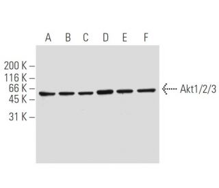
This polyclonal antibody has been discontinued. See our recommended Akt1/2/3 (5C10) monoclonal antibody (sample data shown).
Akt1/2/3 Antibody (H-136): sc-8312
- Akt1/2/3 Antibody (H-136) is a rabbit polyclonal IgG; 200 µg/ml
- Discontinued polyclonal antibody

Ordering Information
Akt1/2/3 (5C10): sc-81434 [ Recommended monoclonal replacement for Akt1/2/3 (H-136) ]
| Product Name | Catalog # | UNIT | Price | Qty | FAVORITES | |
Akt1/2/3 Antibody (5C10) | sc-81434 | 50 µg/0.5 ml | $322.00 | |||
Akt1/2/3 Antibody (5C10): m-IgG Fc BP-HRP Bundle | sc-539612 | 50 µg Ab; 10 µg BP | $361.00 |