")
p-ERK Anticorpo (E-4): sc-7383
- p-ERK Anticorpo E-4é um anticorpo monoclonal produzido em camundongo IgG2a κ, citado em 3.292 publicações, fornecido em 200 µg/ml
- p-ERK Anticorpo (E-4) é recomendado para a detecção de ERK 1 phosphorylated at Tyr 204 and correspondingly phosphorylated ERK 2 de mouse, rat, human e de origem WB, IP, IF, IHC(P) e ELISA; Reage também com outras espécies, incluindo and equine, canine, bovine, porcine and avian
- Anticorpo anti-p-ERK O anticorpo anti-p-ERK (E-4) está disponível conjugado com agarose para IP; HRP para WB, IHC(P) e ELISA; e com fioeritrina ou FITC para IF, IHC(P) e FCM
- também disponível conjugado com Alexa Fluor® 488, Alexa Fluor® 546, Alexa Fluor® 594 ou Alexa Fluor® 647 para WB (RGB), IF, IHC(P) e FCM, e para utilização com sistemas de imagiologia fluorescente RGB, tais como iBright™ FL1000, FluorChem™, Typhoon, Azure e outros sistemas comparáveis
- também disponível conjugado com Alexa Fluor® 680 ou Alexa Fluor® 790 para WB (NIR), IF e FCM; para utilização com sistemas de deteção de infravermelhos próximos (NIR), tais como LI-COR®Odyssey®, iBright™ FL1000, FluorChem™, Typhoon, Azure e outros sistemas comparáveis
- disponível na forma conjugada à biotina para WB, IHC(P) e ELISA; e para TRITC ou Alexa Fluor® 405 para IF, IHC(P) e FCM
- m-IgG Fc BP-HRP e 2a BP-HRP">m-IgG2a BP-HRP são os reagentes de deteção secundários preferidos para p-ERK Antibody (E-4) para aplicações WB e IHC(P). Estes reagentes são agora oferecidos em conjuntos com p-ERK Antibody (E-4)(ver informações de encomenda abaixo).

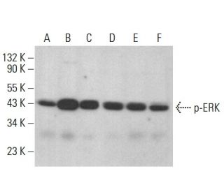
O anticorpo p-ERK (E-4) é um anticorpo monoclonal de cadeia leve IgG2a kappa de ratinho que detecta ERK fosforilada a Tyr 204 em amostras de camundongo, rato e humano através de aplicações como western blotting (WB), imunoprecipitação (IP), imunofluorescência (IF), imunohistoquímica e ensaio imunoenzimático (ELISA). O anticorpo monoclonal p-ERK (E-4) está disponível em formas não conjugadas e em várias formas conjugadas, incluindo agarose, peroxidase de rábano (HRP), ficoeritrina (PE), isotiocianato de fluoresceína (FITC) e vários conjugados Alexa Fluor®. As proteínas ERK, que incluem a ERK 1 e a ERK 2, desempenham um papel crucial na mediação das vias de transdução de sinais desencadeadas por factores de crescimento, hormonas e neurotransmissores. As proteínas ERK estão localizadas no citoplasma e se transloca para o núcleo quando ativadas, onde as proteínas ERK regulam a expressão gênica e influenciam a proliferação, diferenciação e sobrevivência das células. A fosforilação dupla da ERK em resíduos específicos de treonina e tirosina é essencial para a ativação enzimática completa, permitindo a fosforilação de alvos subsequentes em resíduos de serina e treonina. Os principais reguladores ascendentes da ERK incluem a MAP quinase quinase (MEK), a MEK quinase e a Raf-1, que são vitais para o funcionamento correto da cascata de sinalização MAPK. Além disso, a família ERK inclui três outros membros: O anticorpo anti-ERK (E-4) é um recurso inestimável para investigar a fosforilação de ERK em Tyr 204, uma modificação crítica necessária para a ativação e o papel na cascata de sinalização MAPK.
Alexa Fluor® é uma marca comercial da Molecular Probes Inc., OR., EUA
LI-COR® e Odyssey® são marcas registadas da LI-COR Biosciences
Referencias do p-ERK Anticorpo (E-4):
- ERK1 e ERK2, duas proteínas 2 quinases associadas aos microtúbulos, medeiam a fosforilação da tirosina hidroxilase na serina-31 in situ. | Haycock, JW., et al. 1992. Proc Natl Acad Sci U S A. 89: 2365-9. PMID: 1347949
- Purificação de uma proteína-tirosina/treonina quinase murina que fosforila e ativa o produto do gene Erk-1: relação com o produto do gene byr1 da levedura de fissão. | Crews, CM. and Erikson, RL. 1992. Proc Natl Acad Sci U S A. 89: 8205-9. PMID: 1381507
- A estrutura primária da MEK, uma proteína quinase que fosforila o produto do gene ERK. | Crews, CM., et al. 1992. Science. 258: 478-80. PMID: 1411546
- Purificação e propriedades da quinase 1 regulada por sinal extracelular, uma proteína 2 quinase associada a microtúbulos estimulada por insulina. | Boulton, TG., et al. 1991. Biochemistry. 30: 278-86. PMID: 1846291
- Identificação dos locais de fosforilação reguladores na pp42/proteína quinase activada pelo mitogénio (MAP quinase). | Payne, DM., et al. 1991. EMBO J. 10: 885-92. PMID: 1849075
- ERKs: uma família de proteínas-serina/treonina quinases que são activadas e fosforiladas em tirosina em resposta à insulina e ao NGF. | Boulton, TG., et al. 1991. Cell. 65: 663-75. PMID: 2032290
- Componentes de uma nova via de transdução de sinal de proteína quinase humana. | Zhou, G., et al. 1995. J Biol Chem. 270: 12665-9. PMID: 7759517
- ERK6, uma proteína quinase activada por mitogénio envolvida na diferenciação de mioblastos C2C12. | Lechner, C., et al. 1996. Proc Natl Acad Sci U S A. 93: 4355-9. PMID: 8633070
Informacoes sobre ordens
| Nome do Produto | Numero de Catalogo | UNID | Preco | Qde | FAVORITOS | |
p-ERK Anticorpo (E-4) | sc-7383 | 200 µg/ml | $322.00 | |||
Pacote do p-ERK (E-4): m-IgG Fc BP-HRP | sc-525440 | 200 µg Ab; 10 µg BP | $361.00 | |||
Pacote do p-ERK (E-4): m-IgG2a BP-HRP | sc-546255 | 200 µg Ab; 10 µg BP | $361.00 | |||
p-ERK Anticorpo (E-4) AC | sc-7383 AC | 500 µg/ml, 25% agarose | $424.00 | |||
p-ERK Anticorpo (E-4) HRP | sc-7383 HRP | 200 µg/ml | $322.00 | |||
p-ERK Anticorpo (E-4) FITC | sc-7383 FITC | 200 µg/ml | $336.00 | |||
p-ERK Anticorpo (E-4) PE | sc-7383 PE | 200 µg/ml | $349.00 | |||
p-ERK Anticorpo (E-4) Alexa Fluor® 488 | sc-7383 AF488 | 200 µg/ml | $364.00 | |||
p-ERK Anticorpo (E-4) Alexa Fluor® 546 | sc-7383 AF546 | 200 µg/ml | $364.00 | |||
p-ERK Anticorpo (E-4) Alexa Fluor® 594 | sc-7383 AF594 | 200 µg/ml | $364.00 | |||
p-ERK Anticorpo (E-4) Alexa Fluor® 647 | sc-7383 AF647 | 200 µg/ml | $364.00 | |||
p-ERK Anticorpo (E-4) Alexa Fluor® 680 | sc-7383 AF680 | 200 µg/ml | $364.00 | |||
p-ERK Anticorpo (E-4) Alexa Fluor® 790 | sc-7383 AF790 | 200 µg/ml | $364.00 | |||
p-ERK Anticorpo (E-4) B | sc-7383 B | 200 µg/ml | $326.00 | |||
p-ERK Anticorpo (E-4) TRITC | sc-7383 TRITC | 200 µg/ml | $326.00 | |||
p-ERK Anticorpo (E-4) Alexa Fluor® 405 | sc-7383 AF405 | 200 µg/ml | $364.00 | |||
p-ERK (E-4) peptídeo neutralizante | sc-7383 P | 100 µg/0.5 ml | $69.00 |