")
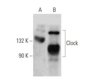
</a> monoclonal antibody (sample data shown).")
This polyclonal antibody has been discontinued. See our recommended Clock (C-8) monoclonal antibody (sample data shown).
Clock Antibody (S-19): sc-6927
- Clock Antibody (S-19) is a goat polyclonal IgG; 200 µg/ml
- epitope mapping at the N-terminus of Clock of mouse origin
- Discontinued polyclonal antibody

SEE ALSO...
Ordering Information
Clock (C-8): sc-271603 [ Recommended monoclonal replacement for Clock (S-19) ]
| Product Name | Catalog # | UNIT | Price | Qty | FAVORITES | |
Clock Antibody (C-8) | sc-271603 | 200 µg/ml | $322.00 | |||
Clock Antibody (C-8): m-IgG2b BP-HRP Bundle | sc-548706 | 200 µg Ab; 10 µg BP | $361.00 | |||
Clock Antibody (C-8) X | sc-271603 X | 200 µg/0.1 ml | $322.00 | |||
Clock Antibody (C-8) AC | sc-271603 AC | 500 µg/ml, 25% agarose | $424.00 | |||
Clock Antibody (C-8) HRP | sc-271603 HRP | 200 µg/ml | $322.00 | |||
Clock Antibody (C-8) FITC | sc-271603 FITC | 200 µg/ml | $336.00 | |||
Clock Antibody (C-8) PE | sc-271603 PE | 200 µg/ml | $349.00 | |||
Clock Antibody (C-8) Alexa Fluor® 488 | sc-271603 AF488 | 200 µg/ml | $364.00 | |||
Clock Antibody (C-8) Alexa Fluor® 546 | sc-271603 AF546 | 200 µg/ml | $364.00 | |||
Clock Antibody (C-8) Alexa Fluor® 594 | sc-271603 AF594 | 200 µg/ml | $364.00 | |||
Clock Antibody (C-8) Alexa Fluor® 647 | sc-271603 AF647 | 200 µg/ml | $364.00 | |||
Clock Antibody (C-8) Alexa Fluor® 680 | sc-271603 AF680 | 200 µg/ml | $364.00 | |||
Clock Antibody (C-8) Alexa Fluor® 790 | sc-271603 AF790 | 200 µg/ml | $364.00 |