")
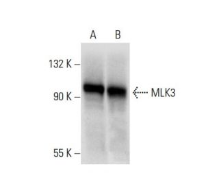
</a> monoclonal antibody (sample data shown).")
This polyclonal antibody has been discontinued. See our recommended MLK3 (D-11) monoclonal antibody (sample data shown).
MLK3 Antibody (C-20): sc-536
- MLK3 Antibody (C-20) is a rabbit polyclonal IgG; 200 µg/ml
- epitope mapping at the C-terminus of MLK3 of human origin
- Discontinued polyclonal antibody

Ordering Information
MLK3 (D-11): sc-166639 [ Recommended monoclonal replacement for MLK3 (C-20) ]
| Product Name | Catalog # | UNIT | Price | Qty | FAVORITES | |
MLK3 Antibody (D-11) | sc-166639 | 200 µg/ml | $322.00 | |||
MLK3 Antibody (D-11): m-IgGκ BP-HRP Bundle | sc-521686 | 200 µg Ab, 40 µg BP | $361.00 | |||
MLK3 Antibody (D-11) AC | sc-166639 AC | 500 µg/ml, 25% agarose | $424.00 | |||
MLK3 Antibody (D-11) HRP | sc-166639 HRP | 200 µg/ml | $322.00 | |||
MLK3 Antibody (D-11) FITC | sc-166639 FITC | 200 µg/ml | $336.00 | |||
MLK3 Antibody (D-11) PE | sc-166639 PE | 200 µg/ml | $349.00 | |||
MLK3 Antibody (D-11) Alexa Fluor® 488 | sc-166639 AF488 | 200 µg/ml | $364.00 | |||
MLK3 Antibody (D-11) Alexa Fluor® 546 | sc-166639 AF546 | 200 µg/ml | $364.00 | |||
MLK3 Antibody (D-11) Alexa Fluor® 594 | sc-166639 AF594 | 200 µg/ml | $364.00 | |||
MLK3 Antibody (D-11) Alexa Fluor® 647 | sc-166639 AF647 | 200 µg/ml | $364.00 | |||
MLK3 Antibody (D-11) Alexa Fluor® 680 | sc-166639 AF680 | 200 µg/ml | $364.00 | |||
MLK3 Antibody (D-11) Alexa Fluor® 790 | sc-166639 AF790 | 200 µg/ml | $364.00 |