")
caveolin-3 Anticorpo (A-3): sc-5310
- caveolin-3 Anticorpo A-3é um anticorpo monoclonal produzido em camundongo IgG1 κ caveolin-3 anticorpo, citado em 65 publicações, fornecido em 200 µg/ml
- específico para um epítopo mapeado entre os aminoácidos3-40 no N-terminus de caveolin-3 de mouse origem
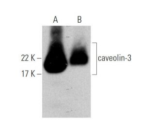
- caveolin-3 Anticorpo (A-3) é recomendado para a detecção de caveolin-3 de mouse, rat e human origem em WB, IP, IF, IHC(P) e ELISA
- Anticorpo anti-caveolin-3 O anticorpo anti-caveolin-3 (A-3) está disponível conjugado com agarose para IP; HRP para WB, IHC(P) e ELISA; e com fioeritrina ou FITC para IF, IHC(P) e FCM
- também disponível conjugado com Alexa Fluor® 488, Alexa Fluor® 546, Alexa Fluor® 594 ou Alexa Fluor® 647 para WB (RGB), IF, IHC(P) e FCM, e para utilização com sistemas de imagiologia fluorescente RGB, tais como iBright™ FL1000, FluorChem™, Typhoon, Azure e outros sistemas comparáveis
- também disponível conjugado com Alexa Fluor® 680 ou Alexa Fluor® 790 para WB (NIR), IF e FCM; para utilização com sistemas de deteção de infravermelhos próximos (NIR), tais como LI-COR®Odyssey®, iBright™ FL1000, FluorChem™, Typhoon, Azure e outros sistemas comparáveis
- m-IgG Fc BP-HRP e m-IgG1 BP-HRP são os reagentes de deteção secundários preferidos para caveolin-3 Antibody (A-3) for WB and IHC(P) applications. Estes reagentes são agora oferecidos em conjuntos com caveolin-3 Antibody (A-3)(ver informações de encomenda abaixo).

LINKS RÁPIDOS
O anticorpo caveolina-3 (A-3) é um anticorpo monoclonal IgG1 de cadeia leve kappa de camundongo que detecta a proteína caveolina-3 de origem camundongo, ratazana e humana por western blotting (WB), imunoprecipitação (IP), imunofluorescência (IF), imunohistoquímica e ensaio imunoenzimático (ELISA). O anticorpo monoclonal caveolina-3 (A-3) está disponível em formas não conjugadas e em várias formas conjugadas, incluindo agarose, peroxidase de rábano (HRP), ficoeritrina (PE), isotiocianato de fluoresceína (FITC) e vários conjugados Alexa Fluor®. A caveolina-3 desempenha um papel crucial na formação de caveolas, que são invaginações especializadas em forma de frasco da membrana plasmática, essenciais para vários processos celulares, incluindo a endocitose e a transdução de sinais. Estas estruturas são particularmente importantes nas células musculares, onde a caveolina-3 é predominantemente expressa, uma vez que facilitam a compartimentação de moléculas de sinalização e a regulação do metabolismo lipídico. A presença de caveolina-3 é vital para manter a integridade das cavéolas e assegurar uma comunicação celular adequada, especialmente em resposta ao stress mecânico e durante a contração muscular. Além disso, a caveolina-3 interage com outras caveolinas e proteínas de sinalização, contribuindo para a regulação dinâmica das vias de sinalização celular. A compreensão da função e da importância da caveolina-3 pode fornecer informações sobre vários processos fisiológicos e potenciais condições patológicas, tornando o anticorpo monoclonal para caveolina-3 (A-3) uma ferramenta inestimável para os investigadores que estudam a biologia muscular e áreas afins.
Alexa Fluor® é uma marca comercial da Molecular Probes Inc., OR., EUA
LI-COR® e Odyssey® são marcas registadas da LI-COR Biosciences
Referencias do caveolin-3 Anticorpo (A-3):
- Caveolina, uma proteína componente das camadas membranares das caveolas. | Rothberg, KG., et al. 1992. Cell. 68: 673-82. PMID: 1739974
- Caveolina-3 e Caveolae regulam a repolarização ventricular. | Markandeya, YS., et al. 2023. J Mol Cell Cardiol. 177: 38-49. PMID: 36842733
- A modificação nitrativa da Caveolina-3: um novo mecanismo de resistência cardíaca à insulina e um potencial alvo terapêutico contra a insuficiência cardíaca isquémica em animais pré-diabéticos. | Meng, Z., et al. 2023. Circulation. 147: 1162-1179. PMID: 36883479
- Caveolin-3 Nitration Drives Insulin Resistance in Prediabetic Hearts. | Sadoshima, J. 2023. Circulation. 147: 1180-1182. PMID: 37036910
- A Interação entre a Caveolina-3 e a Caveolina-1 Diminui a Disfunção dos Canais Devido a Alterações na Caveolina-3. | Benzoni, P., et al. 2024. Int J Mol Sci. 25: PMID: 38256054
- Alterações morfológicas da membrana plasmática de fibroblastos 3T3-L1 após a diferenciação para a forma de adipócito. | Fan, JY., et al. 1983. J Cell Sci. 61: 219-30. PMID: 6885939
- Caracterização de domínios de membrana ricos em caveolina isolados de uma fonte rica em endotelial: implicações para a doença humana. | Lisanti, MP., et al. 1994. J Cell Biol. 126: 111-26. PMID: 7517942
- VIP21/caveolina, aglomerados de glicoesfingolípidos e triagem de proteínas ancoradas no glicosilfosfatidilinositol em células epiteliais. | Zurzolo, C., et al. 1994. EMBO J. 13: 42-53. PMID: 8306971
- Identificação, sequência e expressão de caveolina-2 define uma família de genes de caveolina. | Scherer, PE., et al. 1996. Proc Natl Acad Sci U S A. 93: 131-5. PMID: 8552590
- Clonagem molecular da caveolina-3, um novo membro da família de genes da caveolina expresso predominantemente no músculo. | Tang, Z., et al. 1996. J Biol Chem. 271: 2255-61. PMID: 8567687
- Fosforilação da caveolina por tirosina-quinases src. A isoforma alfa da caveolina é fosforilada seletivamente pela v-Src in vivo. | Li, S., et al. 1996. J Biol Chem. 271: 3863-8. PMID: 8632005
- Expressão da caveolina-3 em células musculares esqueléticas, cardíacas e lisas. A caveolina-3 é um componente do sarcolema e co-fracciona com a distrofina e as glicoproteínas associadas à distrofina. | Song, KS., et al. 1996. J Biol Chem. 271: 15160-5. PMID: 8663016
Informacoes sobre ordens
| Nome do Produto | Numero de Catalogo | UNID | Preco | Qde | FAVORITOS | |
caveolin-3 Anticorpo (A-3) | sc-5310 | 200 µg/ml | $322.00 | |||
Pacote do caveolin-3 (A-3): m-IgG Fc BP-HRP | sc-526483 | 200 µg Ab; 10 µg BP | $361.00 | |||
Pacote do caveolin-3 (A-3): m-IgG1 BP-HRP | sc-531856 | 200 µg Ab; 20 µg BP | $361.00 | |||
caveolin-3 Anticorpo (A-3) AC | sc-5310 AC | 500 µg/ml, 25% agarose | $424.00 | |||
caveolin-3 Anticorpo (A-3) HRP | sc-5310 HRP | 200 µg/ml | $322.00 | |||
caveolin-3 Anticorpo (A-3) FITC | sc-5310 FITC | 200 µg/ml | $336.00 | |||
caveolin-3 Anticorpo (A-3) PE | sc-5310 PE | 200 µg/ml | $349.00 | |||
caveolin-3 Anticorpo (A-3) Alexa Fluor® 488 | sc-5310 AF488 | 200 µg/ml | $364.00 | |||
caveolin-3 Anticorpo (A-3) Alexa Fluor® 546 | sc-5310 AF546 | 200 µg/ml | $364.00 | |||
caveolin-3 Anticorpo (A-3) Alexa Fluor® 594 | sc-5310 AF594 | 200 µg/ml | $364.00 | |||
caveolin-3 Anticorpo (A-3) Alexa Fluor® 647 | sc-5310 AF647 | 200 µg/ml | $364.00 | |||
caveolin-3 Anticorpo (A-3) Alexa Fluor® 680 | sc-5310 AF680 | 200 µg/ml | $364.00 | |||
caveolin-3 Anticorpo (A-3) Alexa Fluor® 790 | sc-5310 AF790 | 200 µg/ml | $364.00 | |||
caveolin-3 (A-3) peptídeo neutralizante | sc-5310 P | 100 µg/0.5 ml | $69.00 |