")
Myc Antibody (C-33): sc-42
- Myc Antibody (C-33) is a mouse monoclonal IgG1 κ, cited in 346 publications, provided at 200 µg/ml
- raised against full length c-Myc of human origin
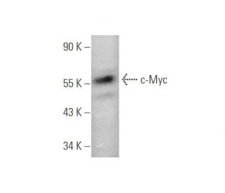
- Anti-Myc Antibody (C-33) is recommended for detection of c-Myc p67 and c-Myc tagged fusion proteins of mouse, rat, human and avian origin by WB, IP, IF and ELISA
- Anti-Myc Antibody (C-33) is available conjugated to agarose for IP; HRP for WB, IHC(P) and ELISA; and to either phycoerythrin or FITC for IF, IHC(P) and FCM
- also available conjugated to Alexa Fluor® 488, Alexa Fluor® 546, Alexa Fluor® 594 or Alexa Fluor® 647 for WB (RGB), IF, IHC(P) and FCM, and for use with RGB fluorescent imaging systems, such as iBright™ FL1000, FluorChem™, Typhoon, Azure and other comparable systems
- also available conjugated to Alexa Fluor® 680 or Alexa Fluor® 790 for WB (NIR), IF and FCM; for use with Near-Infrared (NIR) detection systems, such as LI-COR®Odyssey®, iBright™ FL1000, FluorChem™, Typhoon, Azure and other comparable systems
- TransCruz reagent for Gel Supershift and ChIP applications (sc-42 X, 200 µg/0.1 ml)
- Contact our Technical Service Department (or your local Distributor) for more information on how to receive a FREE 10 µg sample of Myc (C-33): sc-42.
- m-IgG Fc BP-HRP, m-IgG1 BP-HRP and m-IgGκ BP-HRP are the preferred secondary detection reagents for Myc Antibody (C-33) for WB applications. These reagents are now offered in bundles with Myc Antibody (C-33) (see ordering information below).
QUICK LINKS
c-Myc Antibody (C-33) is a mouse monoclonal IgG1 kappa light chain antibody raised against human c-Myc. c-Myc mouse monoclonal antibody (C-33) recognizes an epitope within the c-Myc p67 protein and c-Myc-tagged fusion proteins from mouse, rat, and human sources. Anti-c-Myc monoclonal antibody (C-33) demonstrates high effectiveness in Western blotting, immunoprecipitation, immunofluorescence, and ELISA applications. c-Myc (C-33) antibody is available in non-conjugated form and multiple conjugated forms including agarose, HRP, PE, FITC, and various Alexa Fluor® conjugates, offering flexibility for diverse assay requirements. The c-Myc protein, encoded by the MYC proto-oncogene on chromosome 8q24, functions as a critical transcription factor in cell cycle progression, apoptosis, and cellular transformation. As a member of the Myc family of transcription factors, c-Myc contains several conserved domains, including the basic helix-loop-helix leucine zipper (bHLH-LZ) domain at the C-terminus, essential for DNA binding and dimerization with Max protein. c-Myc regulates numerous target genes involved in ribosome biogenesis, metabolism, protein synthesis, and cell growth. Overexpression or amplification of the c-Myc gene commonly occurs in various human cancers, including Burkitt′s lymphoma, neuroblastoma, and carcinomas of the lung, breast, colon, and cervix. c-Myc deregulation disrupts normal cellular homeostasis, leading to uncontrolled cell proliferation and inhibition of differentiation. Under certain conditions, c-Myc can induce apoptosis as a fail-safe mechanism against tumorigenesis; however, additional oncogenic signals or loss of tumor suppressor functions can circumvent this apoptotic pathway. c-Myc forms heterodimers with Max, another bHLH-LZ protein, to bind specific E-box DNA sequences (CACGTG) in target gene promoters, regulating their transcription. The c-Myc/Max complex acts as a transcriptional activator, while Max can also heterodimerize with other proteins like Mad1 and Mxi1 to form complexes that repress transcription. This dynamic interplay between activation and repression maintains normal cellular functions. Given c-Myc′s central role in cell biology and cancer, c-Myc remains a key target in cancer research.
Alexa Fluor® is a trademark of Molecular Probes Inc., OR., USA
LI-COR® and Odyssey® are registered trademarks of LI-COR Biosciences
Myc Antibody (C-33) References:
- Transcriptional amplification in tumor cells with elevated c-Myc. | Lin, CY., et al. 2012. Cell. 151: 56-67. PMID: 23021215
- c-MYC-induced genomic instability. | Kuzyk, A. and Mai, S. 2014. Cold Spring Harb Perspect Med. 4: a014373. PMID: 24692190
- c-MYC and Epithelial Ovarian Cancer. | Reyes-González, JM. and Vivas-Mejía, PE. 2021. Front Oncol. 11: 601512. PMID: 33718147
- A Selective Small-Molecule c-Myc Degrader Potently Regresses Lethal c-Myc Overexpressing Tumors. | Xu, Y., et al. 2022. Adv Sci (Weinh). 9: e2104344. PMID: 35048559
- c-Myc-IMPDH1/2 axis promotes tumourigenesis by regulating GTP metabolic reprogramming. | Zhang, Q., et al. 2023. Clin Transl Med. 13: e1164. PMID: 36629054
- Cuproptosis engages in c-Myc-mediated breast cancer stemness. | Wang, R., et al. 2023. J Transl Med. 21: 409. PMID: 37353799
- The DUBA-SLC7A11-c-Myc axis is critical for stemness and ferroptosis. | Wang, Z., et al. 2023. Oncogene. 42: 2688-2700. PMID: 37537342
- c-Myc inhibits LAPTM5 expression in B-cell lymphomas. | Zhang, Y., et al. 2023. Ann Hematol. 102: 3499-3513. PMID: 37713124
- USP43 stabilizes c-Myc to promote glycolysis and metastasis in bladder cancer. | Li, M., et al. 2024. Cell Death Dis. 15: 44. PMID: 38218970
- c-Myc alone is enough to reprogram fibroblasts into functional macrophages. | Li, S., et al. 2024. J Hematol Oncol. 17: 83. PMID: 39267119
Ordering Information
| Product Name | Catalog # | UNIT | Price | Qty | FAVORITES | |
Myc Antibody (C-33) | sc-42 | 200 µg/ml | $316.00 | |||
Myc Antibody (C-33): m-IgG Fc BP-HRP Bundle | sc-528091 | 200 µg Ab; 10 µg BP | $354.00 | |||
Myc Antibody (C-33): m-IgGκ BP-HRP Bundle | sc-520389 | 200 µg Ab, 40 µg BP | $354.00 | |||
Myc Antibody (C-33): m-IgG1 BP-HRP Bundle | sc-542755 | 200 µg Ab; 20 µg BP | $354.00 | |||
Myc Antibody (C-33) X | sc-42 X | 200 µg/0.1 ml | $316.00 | |||
Myc Antibody (C-33) AC | sc-42 AC | 500 µg/ml, 25% agarose | $416.00 | |||
Myc Antibody (C-33) HRP | sc-42 HRP | 200 µg/ml | $316.00 | |||
Myc Antibody (C-33) FITC | sc-42 FITC | 200 µg/ml | $330.00 | |||
Myc Antibody (C-33) PE | sc-42 PE | 200 µg/ml | $343.00 | |||
Myc Antibody (C-33) Alexa Fluor® 488 | sc-42 AF488 | 200 µg/ml | $357.00 | |||
Myc Antibody (C-33) Alexa Fluor® 546 | sc-42 AF546 | 200 µg/ml | $357.00 | |||
Myc Antibody (C-33) Alexa Fluor® 594 | sc-42 AF594 | 200 µg/ml | $357.00 | |||
Myc Antibody (C-33) Alexa Fluor® 647 | sc-42 AF647 | 200 µg/ml | $357.00 | |||
Myc Antibody (C-33) Alexa Fluor® 680 | sc-42 AF680 | 200 µg/ml | $357.00 | |||
Myc Antibody (C-33) Alexa Fluor® 790 | sc-42 AF790 | 200 µg/ml | $357.00 |