")
</a>モノクローナル抗体をご覧ください(サンプルデータを表示)。")

注文情報
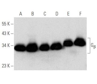
Gβ (H-1): sc-166123 [ Gβ (T-20) の強くお勧めのモノクローナル抗体の代替品]
| 製品名 | カタログ # | 単位 | 価格 | 数量 | お気に入り | |
Gβ 抗体 (H-1) | sc-166123 | 200 µg/ml | $322.00 | |||
Gβ (H-1): m-IgGκ BP-HRP Bundle | sc-521516 | 200 µg Ab, 40 µg BP | $361.00 | |||
Gβ (H-1): m-IgG1 BP-HRP Bundle | sc-533495 | 200 µg Ab; 20 µg BP | $361.00 | |||
Gβ 抗体 (H-1) AC | sc-166123 AC | 500 µg/ml, 25% agarose | $424.00 | |||
Gβ 抗体 (H-1) HRP | sc-166123 HRP | 200 µg/ml | $322.00 | |||
Gβ 抗体 (H-1) FITC | sc-166123 FITC | 200 µg/ml | $336.00 | |||
Gβ 抗体 (H-1) PE | sc-166123 PE | 200 µg/ml | $349.00 | |||
Gβ 抗体 (H-1) Alexa Fluor® 488 | sc-166123 AF488 | 200 µg/ml | $364.00 | |||
Gβ 抗体 (H-1) Alexa Fluor® 546 | sc-166123 AF546 | 200 µg/ml | $364.00 | |||
Gβ 抗体 (H-1) Alexa Fluor® 594 | sc-166123 AF594 | 200 µg/ml | $364.00 | |||
Gβ 抗体 (H-1) Alexa Fluor® 647 | sc-166123 AF647 | 200 µg/ml | $364.00 | |||
Gβ 抗体 (H-1) Alexa Fluor® 680 | sc-166123 AF680 | 200 µg/ml | $364.00 | |||
Gβ 抗体 (H-1) Alexa Fluor® 790 | sc-166123 AF790 | 200 µg/ml | $364.00 | |||
Gβ (H-1) 中和ペプチド | sc-166123 P | 100 µg/0.5 ml | $69.00 |