")
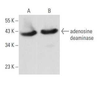
adenosine deaminase Anticuerpo (D-10): sc-376889
- adenosine deaminase Anticuerpo (D-10) es un monoclonal de ratón IgG1 κ, ver las 1 publicaciones, proporcionado como 200 µg/ml
- específico para un epítopo localizado entre los amino ácidos 335-363 en el C-terminus de adenosine deaminase de origen human
- recomendado para detectar adenosine deaminase de human origen, mediante WB, IP, IF, IHC(P) y ELISA
- m-IgG Fc BP-HRP y m-IgG1 BP-HRP son los reactivos de detección secundarios preferidos para adenosine deaminase Anticuerpo (D-10) for WB and IHC(P) applications. Estos reactivos se ofrecen ahora en paquetes con adenosine deaminase Anticuerpo (D-10)(véase la información de pedido más abajo).

ENLACES RÁPIDOS
VER TAMBIÉN ....
Adenosina desaminasa es una enzima que está presente en la mayoría de los tejidos. Existe principalmente como un monómero, aunque en algunos tejidos está asociada con la proteína de unión a la adenosina desaminasa. La adenosina desaminasa degrada la adenosina extracelular, que es tóxica para los linfocitos. La adenosina desaminasa también afecta las señales coestimuladoras en las células T a través de interacciones con CD26. La deficiencia de adenosina desaminasa ha demostrado llevar a enfermedades de inmunodeficiencia como la IDCG (enfermedad de inmunodeficiencia combinada grave) y se ha asociado con anemia hemolítica hereditaria, una enfermedad en la que los niveles de adenosina desaminasa están elevados cincuenta a setenta veces.
Alexa Fluor® es una marca registrada de Molecular Probes Inc., OR., USA
REIVEW LI-COR® y Odyssey® son marcas registradas de LI-COR Biosciences.
adenosine deaminase Anticuerpo (D-10) Referencias:
- Utilización de la adenosina deaminasa y las isoenzimas de la adenosina deaminasa en el diagnóstico de la pleuritis tuberculosa. | Pérez-Rodriguez, E. and Jiménez Castro, D. 2000. Curr Opin Pulm Med. 6: 259-66. PMID: 10912630
- Efectos neuroprotectores de la adenosina deaminasa en el cuerpo estriado. | Tamura, R., et al. 2016. J Cereb Blood Flow Metab. 36: 709-20. PMID: 26746865
- Deficiencia de adenosina deaminasa con una mutación genética novedosa. | Gupta, M., et al. 2016. Indian J Pediatr. 83: 875-6. PMID: 27086606
- La adenosina deaminasa total altamente correlacionada con la actividad de la adenosina deaminasa 2 en suero. | Gao, ZW., et al. 2022. Ann Rheum Dis. 81: e30. PMID: 32001434
- Anomalías urogenitales en la deficiencia de adenosina deaminasa. | Pajno, R., et al. 2020. J Clin Immunol. 40: 610-618. PMID: 32307643
- Adenosina deaminasa - Una diana para nuevos derivados de la piperazina. | Bakaryan, A., et al. 2021. Biophys Chem. 277: 106658. PMID: 34333397
- Variante del gen de la adenosina deaminasa en la diabetes y la obesidad. | Dayani, SB., et al. 2022. J Diabetes Metab Disord. 21: 333-338. PMID: 35673471
- Displasia esquelética en la deficiencia de adenosina deaminasa. | Minagawa, H., et al. 2022. Pediatr Int. 64: e15214. PMID: 35791036
- Directrices actualizadas para el tratamiento del déficit de adenosina deaminasa. | Grunebaum, E., et al. 2023. J Allergy Clin Immunol Pract. 11: 1665-1675. PMID: 36736952
- Las isoenzimas de la adenosina deaminasa y la tuberculosis pleural. | Antony, VB. 1996. J Lab Clin Med. 127: 326-7. PMID: 8656033
Información sobre pedidos
| Nombre del producto | Número de catálogo | UNIDAD | Precio | CANTIDAD | Favoritos | |
adenosine deaminase Anticuerpo (D-10) | sc-376889 | 200 µg/ml | $322.00 | |||
Paquete de adenosine deaminase (D-10): m-IgG Fc BP-HRP | sc-540661 | 200 µg Ab; 10 µg BP | $361.00 | |||
Paquete de adenosine deaminase (D-10): m-IgG1 BP-HRP | sc-542278 | 200 µg Ab; 20 µg BP | $361.00 | |||
adenosine deaminase (D-10) péptido neutralizante | sc-376889 P | 100 µg/0.5 ml | $69.00 |