")
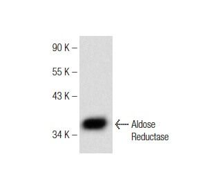
: sc-373953. Western blot analysis of Aldose Reductase expression in Caki-1 whole cell lysate.")

Aldose Reductase Antibody (E-12): sc-373953
- Aldose Reductase Antibody (E-12) is a mouse monoclonal IgG2a κ, cited in 1 publications, provided at 200 µg/ml
- specific for an epitope mapping between amino acids 132-159 within an internal region of Aldose Reductase of human origin
- recommended for detection of Aldose Reductase of human origin by WB, IP, IF, IHC(P) and ELISA
- m-IgG Fc BP-HRP, m-IgG2a BP-HRP and m-IgGκ BP-HRP are the preferred secondary detection reagents for Aldose Reductase Antibody (E-12) for WB and IHC(P) applications. These reagents are now offered in bundles with Aldose Reductase Antibody (E-12) (see ordering information below).

QUICK LINKS
SEE ALSO...
Aldose Reductase Antibody (E-12) is a mouse monoclonal IgG2a antibody that detects Aldose Reductase in human samples through various applications including western blotting (WB), immunoprecipitation (IP), immunofluorescence (IF), immunohistochemistry with paraffin-embedded sections (IHCP), and enzyme-linked immunosorbent assay (ELISA). Aldose Reductase plays a crucial role in cellular metabolism by catalyzing the reduction of aldehydes, particularly converting glucose into sorbitol, which is significant in the context of diabetic complications. While this pathway is generally minor in glucose metabolism across most tissues, excessive sorbitol accumulation during diabetic hyperglycemia leads to hyperosmotic stress on cells. This stress contributes to various diabetic complications, including neuropathy, retinopathy, and cataracts, highlighting Aldose Reductase′s importance in understanding and potentially mitigating these conditions. Aldose Reductase shares structural similarities with human aldehyde reductase (ALR1), bovine prostaglandin F synthase, and the European common frog protein, rho-crystallin, underscoring evolutionary significance and functional similarities across species.
Ordering Information
| Product Name | Catalog # | UNIT | Price | Qty | FAVORITES | |
Aldose Reductase Antibody (E-12) | sc-373953 | 200 µg/ml | $322.00 | |||
Aldose Reductase Antibody (E-12): m-IgG Fc BP-HRP Bundle | sc-537892 | 200 µg Ab; 10 µg BP | $361.00 | |||
Aldose Reductase Antibody (E-12): m-IgGκ BP-HRP Bundle | sc-535348 | 200 µg Ab; 40 µg BP | $361.00 | |||
Aldose Reductase Antibody (E-12): m-IgG2a BP-HRP Bundle | sc-548139 | 200 µg Ab; 10 µg BP | $361.00 | |||
Aldose Reductase (E-12) Neutralizing Peptide | sc-373953 P | 100 µg/0.5 ml | $69.00 |