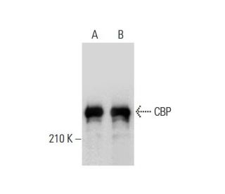
 |
- rabbit polyclonal IgG, 200 µg/ml
- epitope mapping at the N-terminus of CBP of human origin
- recommended for detection of CBP p265 of mouse, rat and human origin by WB, IP, IF and ELISA; also reactive with additional species, including canine, bovine and porcine
- TransCruz reagent for Gel Supershift and ChIP applications, sc-369 X, 200 µg/0.1 ml
- blocking peptide, sc-369 P
|
|
| |
Ordering InformationProduct Citations
Recommended Support Products:
(click button of application of choice)
| |
| Species |
Gene Name |
Gene ID |
Chromosome Location |
Isoform (mRNA) Accession # |
Protein Accession # |
OMIM™ Number |
| Human |
CREBBP |
1387 |
16p13.3 |
NM_004380 |
Q92793
|
600140 |
| Mouse |
Crebbp |
12914 |
16 A1 |
|
P45481
|
N/A |
| |
CBP Background Information Cyclic AMP-regulated gene expression frequently involves a DNA element designated the cAMP-regulated enhancer (CRE). Many transcription factors bind to this element, including the protein CREB, which is activated as a result of phosphorylation by protein kinase A. It has been shown that protein kinase A-mediated CREB phosphorylation results in its binding to a nuclear protein designated CBP (for CREB-binding protein). These findings suggest that CBP has many of the properties expected of a CREB co-activator. Another high molecular weight transcriptional adapter protein, designated p300, is characterized by three cysteine- and histidine-rich regions, of which the most carboxy terminal region specifically binds the adenovirus E1A protein. p300 molecules lacking an intact E1A binding site bypass E1A repression, even in the presence of high concentrations of E1A. Sequence analysis of CBP and p300 has revealed substantial homology, arguing that these proteins are members of a conserved family of co-activators.
| CBP (A-22) Product Citations |
See how others have used CBP (A-22): sc-369 antibody and or CBP (A-22) antibody conjugates.
263 total citations Loading citations.
|
| |
|
 |
|
CBP (A-22)
Click on image to enlarge
|
|
CBP (A-22): sc-369. Western blot analysis of CBP expression in KNRK nuclear extract.
ChIP analysis of transcription factor binding to the Interferon β promoter before (A) and six hours after (B) Sendai virus infection of HeLa cells. Antibodies tested included NFκB p65 (A): sc-109, ATF-2 (N-96): sc-6233, GCN5 (N-18): sc-6303, Pol II (H-224): sc-9001, TFIID (TBP)(SI-1): sc-273 and CBP (A-22): sc-369. Data kindly provided by G. Mosialos.
ChIP analysis of coactivator recruitment on Cyclin D2 promoter in C2C12 cells treated with LiCl and serum. Antibodies tested include β-catenin (H-102): sc-7199, β-catenin (C-18): sc-1496, β-catenin (E-5): sc-7963, Tip60 (N-17): sc-5725, TRRAP (T-17): sc-5405, TRRAP (Y-18): sc-12375, TRRAP (F-20): sc-12376, TRRAP (H-300): sc-11411, CBP (A-22): sc-369, CBP (C-20): sc-583, CBP (451): sc-1211, CPB (C-1): sc-7300, p300 (H-272): sc-8981, p300 (N-15): sc-584 and p300 (C-20): sc-585. Data kindly provided by M.G. Rosenfeld and reproduced with permission from Kioussi et al., Cell 2002, 111: 673-685.
ChIP analysis of p52 promoter occupancy in murine p19 cells in response to activation by estradiol (E2). Antibodies tested include ERα (MC-20): sc-542, ERα (H-184): sc-7207, CBP (A-22): sc-369, CBP (C-1): sc-7300, p/CIP (F2): sc-5305, p/CIP (M-397): sc-9119, RIP140 (H-300): sc-8997 and HDAC3 (N-19): sc-11417. Data kindly provided by M.G. Rosenfeld and reproduced with permission from Perissi et al., Cell 2004, 116: 511-526.
ChIP analysis of cofactor occupancy dynamics on the ICAM1 promoter in 293 cells in response to IL-1β treatment. Antibodies tested include NFκB p50 (C-19): sc-1190, NFκB p50 (E-10): sc-8414, NFκB p50 (H-119): sc-7178, NFκB p65 (C-20): sc-372, NFκB p65 (A): sc-109, NFκB p65 (H-286): sc-7151, Bcl-3 (C-14): sc-185, Bcl-3 (H-146): sc-13038, PCAF (C-16): sc-6300, PCAF (H-369): sc-8999, CBP (A-22): sc-369, CBP (C-1): sc-7300, CBP (C-20): sc-583, CBP (451): sc-1211. Data kindly provided by M.G. Rosenfeld and reproduced with permission from Baek et al., Cell 2002, 110: 55-67.
ChIP analysis of cofactor occupancy dynamics on the IL-8 promoter in 293 cells in response to IL-1β treatment. Antibodies tested include NFκB p50 (C-19): sc-1190, NFκB p50 (E-10): sc-8414, NFκB p50 (H-119): sc-7178, NFκB p65 (C-20): sc-372, NFκB p65 (A): sc-109, NFκB p65 (H-286): sc-7151, CBP (A-22): sc-369, CBP (C-1): sc-7300, CBP (C-20): sc-583, CBP (451): sc-1211, p300 (C-20): sc- sc-585, p300 (N-15): sc-584, p300 (H-272): sc-8981. Data kindly provided by M.G. Rosenfeld and reproduced with permission from Baek et al., Cell 2002, 110: 55-67.
ChIP analysis of in vivo binding of RORα and its recruitment of coactivators to RORα-responsive promoters in freshly dissected cerebella derived from wild type (+/+) and staggerer (Sg) mice. Control Input (A). Antibodies used included RORα (C-16): sc-6062 (B), TIP60 (N-17): sc-5725 (C), CBP (A-22): sc-369 and CBP (C-20): sc-583 (D). Data kindly provided by M.G. Rosenfeld and reproduced wtih permission from Gold et al., Neuron 2003, 40: 1119-1131.
ChIP analysis of recruitment of CBP, p300, PCA and BRG1 to the IL-2α (CD25) promoter in vivo. Antibodies tested include CBP (A-22): sc-369, p300 (N-15): sc-584, PCAF (H-369): sc-8999, and Brg-1 (H-88): sc-10768. CD3ε and Oct-2 promoter regions were employed as positive and negative controls, respectively. DNA was isolated from Rag -/- thymocytes or Rag -/- thymocytes expressing TCRβ. Data kindly provided by J. Imbert and reproduced from Yeh, J-H., et al., Nucleic Acids Research, 2002, 30: 1944-1951, with permission from Oxford University Press.
|
|
Download
|
|