")
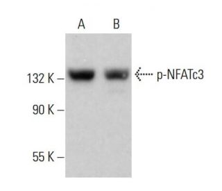
: sc-365786. Jurkat (A) 和 KNRK (B) 核提取物中 NFATc3 磷酸化的 Western 印迹分析.")
 和λ蛋白磷酸酶 (sc-200312A) 处理 (C, F) 的 Jurkat 全细胞裂解液 (A, D) 和 Jurkat 核提取物 (B, C, E, F) 中 NFATc3 磷酸化的 Western 印迹分析. 测试的抗体包括 p-NFATc3 抗体 (C-3): sc-365786 (A, B, C) 和 NFATc3 (F-1): sc-8405 (D, E, F).")
: sc-365786. PC-12 全细胞裂解液中 p-NFATc3 表达的 Western 印迹分析.")
p-NFATc3 抗体 (C-3): sc-365786. Jurkat (A) 和 KNRK (B) 核提取物中 NFATc3 磷酸化的 Western 印迹分析.
p-NFATc3 抗体 (C-3): sc-365786
- p-NFATc3抗体(C-3)是小鼠单克隆IgG1 κ, 在4篇文献中引用,规格为200 µg/ml
- 抗-p-NFATc3 抗体 (C-3) 推荐用于 WB, IP, IF 和 ELISA,检测mouse, rat 和human 来源的 Ser 240 phosphorylated NFATc3
- 抗p-NFATc3抗体(C-3)可与琼脂糖结合用于IP;与HRP结合用于WB、IHC(P)和ELISA;与藻红蛋白或FITC结合用于IF、IHC(P)和FCM
- 还可偶联Alexa Fluor® 488, Alexa Fluor® 546, Alexa Fluor® 594 和 Alexa Fluor® 647,用于WB (RGB), IF, IHC(P) 和 FCM, 以及用于RGB荧光成像系统,例如iBright™ FL1000, FluorChem™, Typhoon, Azure和其他类似的系统
- 还可偶联Alexa Fluor® 680 和 Alexa Fluor® 790, 用于WB (NIR), IF 和 FCM; 以及用于近红外(NIR)检测系统,如LI-COR®/Odyssey®, iBright™ FL1000, FluorChem™, Typhoon, Azure和类似系统
- m-IgG Fc BP-HRP和m-IgGκ BP-HRP是p-NFATc3 Antibody (C-3) 适用于 WB 应用。 的首选辅助检测试剂,这些试剂现与p-NFATc3 Antibody (C-3) 打包提供(请参阅下面的订购信息)。

p-NFATc3抗体(C-3)是一种IgG1 κ小鼠单克隆p-NFATc3抗体,能够通过WB、IP、IF和ELISA等方法检测小鼠、大鼠和人类来源的Ser 240磷酸化NFATc3。p-NFATc3抗体(C-3)既有非偶联形式的抗p-NFATc3抗体,也有多种偶联形式的抗p-NFATc3抗体,包括琼脂糖、HRP、PE、FITC和多种Alexa Fluor®偶联物。NFAT(活化T细胞核因子)转录因子家族的成员与NFκB/Rel蛋白有关,并在DNA上与AP-1蛋白(Fos和Jun)形成协作复合物,以调节T细胞中的细胞因子表达。NFAT蛋白广泛表达并经过选择性修饰以产生剪接变体,它们既定位于细胞质(NFATc)也定位于细胞核(NFATn)。NFATc1(NFATc)、NFATc2(NFATp)和NFATc3(NFAT4,NFSTx)主要在免疫细胞中表达。NFAT蛋白由细胞内钙的增加而激活,这导致钙调蛋白依赖性磷酸酶--钙调神经磷酸酶使NFAT蛋白去磷酸化。这种激活事件诱导蛋白质结构发生构象变化,暴露出核定位信号,并促进NFAT蛋白从细胞质向细胞核的易位。
仅限研究使用。不适用于诊断和治疗用途。
Alexa Fluor® 是Molecular Probes Inc., OR., USA的商标
LI-COR®和 Odyssey® 是LI-COR Biosciences的注册商标
p-NFATc3 抗体 (C-3) 参考文献:
- 识别免疫细胞激活时诱导基因中的 NFATp/AP-1 复合元素。 | Kel, A., et al. 1999. J Mol Biol. 288: 353-76. PMID: 10329147
- NFAT5 是一种组成型核 NFAT 蛋白,它不与 Fos 和 Jun 合作。 | Lopez-Rodríguez, C., et al. 1999. Proc Natl Acad Sci U S A. 96: 7214-9. PMID: 10377394
- NFATc1 核占位增强会导致 T 细胞活化,而与 CD28 成本刺激无关。 | Pan, M., et al. 2007. J Immunol. 178: 4315-21. PMID: 17371988
- 缺氧诱导的NFATc3去SUMOyl化会促进胰腺癌的进展。 | Tong, Y., et al. 2022. Cell Death Dis. 13: 413. PMID: 35484132
- NF-AT 基因家族两个新成员的分离及 NF-AT 蛋白的功能特征。 | Hoey, T., et al. 1995. Immunity. 2: 461-72. PMID: 7749981
- 活化 T 细胞核因子(NFAT)基因家族成员 NFATc 新异构体的特征。 | Park, J., et al. 1996. J Biol Chem. 271: 20914-21. PMID: 8702849
- 正常人 T 细胞中 NFAT 家族蛋白的表达。 | Lyakh, L., et al. 1997. Mol Cell Biol. 17: 2475-84. PMID: 9111316
- NFAT 家族的转录因子:调节和功能。 | Rao, A., et al. 1997. Annu Rev Immunol. 15: 707-47. PMID: 9143705
- 不同的 NFAT 家族蛋白参与了人类胸腺细胞亚群的核 NFAT-DNA 结合复合物。 | Amasaki, Y., et al. 1998. J Immunol. 160: 2324-33. PMID: 9498773
- 转录因子 NF-ATc 对心脏瓣膜的形成至关重要。 | Ranger, AM., et al. 1998. Nature. 392: 186-90. PMID: 9515964
订购信息
| 产品名称 | 产品编号 | 规格 | 价格 | 数量 | 收藏夹 | |
p-NFATc3 抗体 (C-3) | sc-365786 | 200 µg/ml | $322.00 | |||
p-NFATc3 (C-3): m-IgG Fc BP-HRP 套装 | sc-529466 | 200 µg Ab; 10 µg BP | $361.00 | |||
p-NFATc3 (C-3): m-IgGκ BP-HRP 套装 | sc-522367 | 200 µg Ab, 40 µg BP | $361.00 | |||
p-NFATc3 抗体 (C-3) AC | sc-365786 AC | 500 µg/ml, 25% agarose | $424.00 | |||
p-NFATc3 抗体 (C-3) HRP | sc-365786 HRP | 200 µg/ml | $322.00 | |||
p-NFATc3 抗体 (C-3) FITC | sc-365786 FITC | 200 µg/ml | $336.00 | |||
p-NFATc3 抗体 (C-3) PE | sc-365786 PE | 200 µg/ml | $349.00 | |||
p-NFATc3 抗体 (C-3) Alexa Fluor® 488 | sc-365786 AF488 | 200 µg/ml | $364.00 | |||
p-NFATc3 抗体 (C-3) Alexa Fluor® 546 | sc-365786 AF546 | 200 µg/ml | $364.00 | |||
p-NFATc3 抗体 (C-3) Alexa Fluor® 594 | sc-365786 AF594 | 200 µg/ml | $364.00 | |||
p-NFATc3 抗体 (C-3) Alexa Fluor® 647 | sc-365786 AF647 | 200 µg/ml | $364.00 | |||
p-NFATc3 抗体 (C-3) Alexa Fluor® 680 | sc-365786 AF680 | 200 µg/ml | $364.00 | |||
p-NFATc3 抗体 (C-3) Alexa Fluor® 790 | sc-365786 AF790 | 200 µg/ml | $364.00 | |||
p-NFATc3 (C-3) 中和勝肽 | sc-365786 P | 100 µg/0.5 ml | $69.00 |