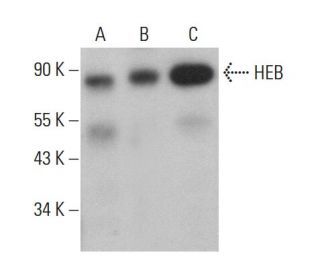
 |
- rabbit polyclonal IgG, 200 µg/ml
- epitope mapping within the C-terminus of HEB of human origin
- recommended for detection of HEB of mouse, rat and human origin by WB, IP, IF, ELISA and indirect FCM using PE-conjugated goat anti-rabbit IgG: sc-3739; also reactive with additional species, including equine, canine, bovine, porcine and avian
- blocking peptide, sc-357 P
- TransCruz reagent for Gel Supershift and ChIP applications, sc-357 X, 200 µg/0.1 ml
|
|
| |
Ordering InformationProduct Citations
Recommended Support Products:
(click button of application of choice)
HEB Background Information Differentiation of myogenic cells is regulated by multiple positively and negatively acting factors. One well characterized family of helix-loop-helix (HLH) proteins known to play an important role in the regulation of muscle cell development includes Myo D, myogenin, Myf-5 and Myf-6 (also designated MRF-4 or herculin). Myo D transcription factors form heterodimers with products of a more widely expressed family of bHLH genes, the E family, which consists of at least three distinct genes: E2A, IF2 and HEB. Myo D-E heterodimers bind avidly to consensus (CANNTG) E box target sites that are functionally important elements in the upstream regulatory sequences of many muscle-specific terminal differentiation genes. Both homo- and hetero-oligomers of these proteins are able to distinguish very closely related E box proteins and are believed to play important roles in lineage specific gene expression.
| HEB (A-20) Product Citations |
See how others have used HEB (A-20): sc-357 antibody and or HEB (A-20) antibody conjugates.
19 total citations Loading citations.
|
| |
|
 |
|
HEB (A-20)
Click on image to enlarge
|
|
HEB (A-20): sc-357. Western blot analysis of HEB expression in Jurkat nuclear extract.
HEB (A-20): sc-357. Immunofluorescence staining of methanol-fixed Jurkat cells showing nuclear staining.
HEB (A-20): sc-357. Indirect, intracellular FCM analysis of fixed and permeabilized Jurkat cells stained with HEB (A-20), followed by PE-conjugated goat anti-rabbit IgG: sc-3739. Black line histogram represents the isotype control, normal rabbit IgG: sc-3888.
HEB (A-20): sc-357. Western blot analysis of HEB expression in non-transfected 293T: sc-117752 (A), human HEB transfected 293T: sc-116193 (B) and Jurkat (C) whole cell lysates.
HEB (A-20): sc-357. Western blot analysis of HEB expression in 293T (A) and SW480 (B) whole cell lysates.
HEB (A-20): sc-357. Indirect, intracellular FCM analysis of fixed and permeabilized Jurkat cells stained with HEB (A-20), followed by FITC-conjugated bovine anti-rabbit IgG: sc-2365. Black line histogram represents the isotype control, normal rabbit IgG: sc-3888.
HEB (A-20): sc-357. Indirect, intracellular FCM analysis of fixed and permeabilized Jurkat cells stained with HEB (A-20), followed by PerCP-conjugated goat anti-rabbit IgG: sc-45090. Black line histogram represents the isotype control, normal rabbit IgG: sc-3888.
HEB (A-20): sc-357. Indirect, intracellular FCM analysis of fixed and permeabilized Jurkat cells stained with HEB (A-20), followed by FITC-conjugated mouse anti-rabbit IgG: sc-2359. Black line histogram represents the isotype control, normal rabbit IgG: sc-3888.
HEB (A-20): sc-357. Indirect, intracellular FCM analysis of fixed and permeabilized Jurkat cells stained with HEB (A-20), followed by PerCP-conjugated mouse anti-rabbit IgG: sc-45098. Black line histogram represents the isotype control, normal rabbit IgG: sc-3888.
|
|
Download
|
|