")
</a>モノクローナル抗体をご覧ください(サンプルデータを表示)。")
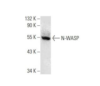
このポリクローナル抗体は販売終了となりました。弊社推奨のN-WASP (C-1)モノクローナル抗体をご覧ください(サンプルデータを表示)。
N-WASP抗体(H-100): sc-20770
- N-WASP抗体 H-100はウサギポリクローナルIgGです。200 µg/mlで提供
- エピトープはhuman由来のN-WASPの内部領域に位置するアミノ酸111-210に対応します
- 生産中止となったポリクローナル抗体

関連項目
注文情報
N-WASP (C-1): sc-271484 [ N-WASP (H-100) の強くお勧めのモノクローナル抗体の代替品]
| 製品名 | カタログ # | 単位 | 価格 | 数量 | お気に入り | |
N-WASP 抗体 (C-1) | sc-271484 | 200 µg/ml | $322.00 | |||
N-WASP (C-1): m-IgG Fc BP-HRP Bundle | sc-529193 | 200 µg Ab; 10 µg BP | $361.00 | |||
N-WASP (C-1): m-IgGκ BP-HRP Bundle | sc-521957 | 200 µg Ab, 40 µg BP | $361.00 | |||
N-WASP 抗体 (C-1) AC | sc-271484 AC | 500 µg/ml, 25% agarose | $424.00 | |||
N-WASP 抗体 (C-1) HRP | sc-271484 HRP | 200 µg/ml | $322.00 | |||
N-WASP 抗体 (C-1) FITC | sc-271484 FITC | 200 µg/ml | $336.00 | |||
N-WASP 抗体 (C-1) PE | sc-271484 PE | 200 µg/ml | $349.00 | |||
N-WASP 抗体 (C-1) Alexa Fluor® 488 | sc-271484 AF488 | 200 µg/ml | $364.00 | |||
N-WASP 抗体 (C-1) Alexa Fluor® 546 | sc-271484 AF546 | 200 µg/ml | $364.00 | |||
N-WASP 抗体 (C-1) Alexa Fluor® 594 | sc-271484 AF594 | 200 µg/ml | $364.00 | |||
N-WASP 抗体 (C-1) Alexa Fluor® 647 | sc-271484 AF647 | 200 µg/ml | $364.00 | |||
N-WASP 抗体 (C-1) Alexa Fluor® 680 | sc-271484 AF680 | 200 µg/ml | $364.00 | |||
N-WASP 抗体 (C-1) Alexa Fluor® 790 | sc-271484 AF790 | 200 µg/ml | $364.00 | |||
N-WASP (C-1) 中和ペプチド | sc-271484 P | 100 µg/0.5 ml | $69.00 |