")
CD44 Anticuerpo (IM7): sc-18849
- CD44 Anticuerpo IM7 es un monoclonal de rata IgG2b, ver las 48 publicaciones, proporcionado 200 µg/ml
- criado contra células inducidas por dexametasona de la leucemia mieloide espontánea M1 de SJL mouse.
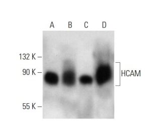
- CD44 Anticuerpo (IM7) es recomendado para detectar HCAM de mouse, rat, human, canine, feline y equine origen, mediante WB, IP, IF, IHC(P) y FCM
- CD44 Anticuerpo (IM7) es disponible conjugado a agarosa para IP; HRP para WB, IHC(P) y ELISA; y tanto a phycoerythrin como a FITC para IF, IHC(P) y FCM
- también disponible conjugado a Alexa Fluor® 488, Alexa Fluor® 546, Alexa Fluor® 594 o Alexa Fluor® 647 para WB (RGB), IF, IHC (P) y FCM
- también disponible conjugado a Alexa Fluor® 680 o Alexa Fluor® 790 para WB (NIR), IF y FCM
- Actualmente, aún no hemos completado la identificación de los reactivos de detección secundaria preferidos para CD44 Anticuerpo (IM7). Este trabajo está en progreso.

ENLACES RÁPIDOS
VER TAMBIÉN ....
El anticuerpo anti-HCAM (IM7) es un anticuerpo monoclonal IgG2b de rata que detecta HCAM en muestras de ratón, rata, humano, canino, felino y equino a través de aplicaciones como western blotting (WB), inmunoprecipitación (IP), inmunofluorescencia (IF), inmunohistoquímica con secciones incluidas en parafina (IHCP) y citometría de flujo (FCM). El anticuerpo anti-HCAM (IM7) está disponible tanto en forma no conjugada como en varias formas conjugadas, incluyendo agarosa, peroxidasa de rábano picante (HRP), ficoeritrina (PE), isotiocianato de fluoresceína (FITC) y múltiples conjugados Alexa Fluor®. La HCAM, también conocida como CD44, es una proteína de membrana de tipo I de paso único de 742 aminoácidos que desempeña un papel crucial en la adhesión y migración celular, funcionando como receptor del ácido hialurónico (AH) e interactuando con ligandos como la osteopontina (OPN). Esta interacción media las conexiones célula-célula y célula-matriz, esenciales durante procesos como la hematopoyesis, la activación de linfocitos y la metástasis tumoral. La estructura de la HCAM incluye un dominio de enlace y sufre empalmes alternativos, lo que da lugar a múltiples isoformas, entre ellas CD44R, CDw44, CD44S, CD44H (hematopoyética) y CD44E (epitelial). La isoforma CD44H muestra una elevada expresión en los tejidos cancerosos, lo que indica un papel significativo en la progresión tumoral y pone de relieve la importancia de la HCAM tanto en los procesos fisiológicos normales como en las condiciones patológicas.
Alexa Fluor® es una marca registrada de Molecular Probes Inc., OR., USA
REIVEW LI-COR® y Odyssey® son marcas registradas de LI-COR Biosciences.
Información sobre pedidos
| Nombre del producto | Número de catálogo | UNIDAD | Precio | CANTIDAD | Favoritos | |
CD44 Anticuerpo (IM7) | sc-18849 | 200 µg/ml | $322.00 | |||
CD44 Anticuerpo (IM7) AC | sc-18849 AC | 500 µg/ml, 25% agarose | $424.00 | |||
CD44 Anticuerpo (IM7) HRP | sc-18849 HRP | 200 µg/ml | $322.00 | |||
CD44 Anticuerpo (IM7) FITC | sc-18849 FITC | 200 µg/ml | $336.00 | |||
CD44 Anticuerpo (IM7) PE | sc-18849 PE | 200 µg/ml | $349.00 | |||
CD44 Anticuerpo (IM7) Alexa Fluor® 488 | sc-18849 AF488 | 200 µg/ml | $364.00 | |||
CD44 Anticuerpo (IM7) Alexa Fluor® 546 | sc-18849 AF546 | 200 µg/ml | $364.00 | |||
CD44 Anticuerpo (IM7) Alexa Fluor® 594 | sc-18849 AF594 | 200 µg/ml | $364.00 | |||
CD44 Anticuerpo (IM7) Alexa Fluor® 647 | sc-18849 AF647 | 200 µg/ml | $364.00 | |||
CD44 Anticuerpo (IM7) Alexa Fluor® 680 | sc-18849 AF680 | 200 µg/ml | $364.00 | |||
CD44 Anticuerpo (IM7) Alexa Fluor® 790 | sc-18849 AF790 | 200 µg/ml | $364.00 |