")
NFATc1 Antibody (H-10): sc-17834
- NFATc1 Antibody (H-10) is a mouse monoclonal IgG1 κ, cited in 18 publications, provided at 200 µg/ml
- raised against amino acids 1-110 of NFATc1 of human origin
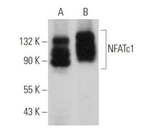
- recommended for detection of NFATc1 of mouse, rat and human origin by WB, IP, IF, IHC(P) and ELISA
- TransCruz reagent for Gel Supershift and ChIP applications (sc-17834 X, 200 µg/0.1 ml)
- See NFATc1 (7A6): sc-7294 for NFATc1 antibody conjugates, including AC, HRP, FITC, PE, Alexa Fluor® 488, 594, 647, 680 and 790.
- m-IgGκ BP-HRP is the preferred secondary detection reagent for NFATc1 Antibody (H-10) for WB and IHC(P) applications. This reagent is now offered in a bundle with NFATc1 Antibody (H-10) (see ordering information below). For additional m-IgGκ BP conjugates see our complete list of Mouse IgG Binding Proteins.

QUICK LINKS
NFATc1 Antibody (H-10) is a mouse monoclonal IgG1 kappa antibody raised against amino acids 1-110 of human NFATc1, also known as Nuclear Factor of Activated T-cells cytoplasmic 1 or NFAT2. NFATc1 is a transcription factor that plays a pivotal role in the immune system by regulating gene expression during T-cell activation and differentiation. Upon activation by calcium signaling, NFATc1 is dephosphorylated by the phosphatase calcineurin, allowing NFATc1 to translocate from the cytoplasm to the nucleus; this nuclear localization enables NFATc1 to bind DNA and modulate the transcription of genes involved in immune responses. The proper function of NFATc1 is essential for cytokine production and the development of effective immune reactions. Dysregulation of NFATc1 activity has been implicated in various pathological conditions, including autoimmune diseases and cancers. Anti-NFATc1 antibody (H-10) reacts with mouse, rat, and human NFATc1 and is suitable for applications such as western blotting (WB), immunoprecipitation (IP), immunofluorescence (IF), immunohistochemistry with paraffin-embedded sections (IHCP), and enzyme-linked immunosorbent assay (ELISA). By targeting NFATc1, anti-NFATc1 antibody (H-10) assists researchers in studying the molecular mechanisms of T-cell function and the broader implications in immunological disorders. Understanding the localization and function of NFATc1 is important for developing therapeutic strategies against diseases resulting from immune system dysregulation.
Ordering Information
| Product Name | Catalog # | UNIT | Price | Qty | FAVORITES | |
NFATc1 Antibody (H-10) | sc-17834 | 200 µg/ml | $322.00 | |||
NFATc1 Antibody (H-10): m-IgGκ BP-HRP Bundle | sc-533747 | 200 µg Ab; 40 µg BP | $361.00 | |||
NFATc1 Antibody (H-10) X | sc-17834 X | 200 µg/0.1 ml | $322.00 |