")
Syk Antibody (4D10): sc-1240
- Syk Antibody (4D10) is a mouse monoclonal IgG2a κ Syk antibody, cited in 269 publications, provided at 200 µg/ml
- raised against amino acids 313-339 of Syk of human origin
- Syk Antibody (4D10) is recommended for detection of Syk of mouse, rat and human origin by WB, IP, IF, IHC(P) and FCM
- Anti-Syk Antibody (4D10) is available conjugated to agarose for IP; HRP for WB, IHC(P) and ELISA; and to either phycoerythrin or FITC for IF, IHC(P) and FCM
- also available conjugated to Alexa Fluor® 488, Alexa Fluor® 546, Alexa Fluor® 594 or Alexa Fluor® 647 for WB (RGB), IF, IHC(P) and FCM, and for use with RGB fluorescent imaging systems, such as iBright™ FL1000, FluorChem™, Typhoon, Azure and other comparable systems
- also available conjugated to Alexa Fluor® 680 or Alexa Fluor® 790 for WB (NIR), IF and FCM; for use with Near-Infrared (NIR) detection systems, such as LI-COR®Odyssey®, iBright™ FL1000, FluorChem™, Typhoon, Azure and other comparable systems
- m-IgG Fc BP-HRP, m-IgG2a BP-HRP and m-IgGκ BP-HRP are the preferred secondary detection reagents for Syk Antibody (4D10) for WB and IHC(P) applications. These reagents are now offered in bundles with Syk Antibody (4D10) (see ordering information below).

QUICK LINKS
SEE ALSO...
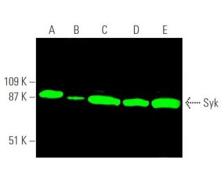
Syk Antibody (4D10) is a mouse monoclonal IgG2a kappa light chain antibody that detects Syk protein of mouse, rat, and human origin by western blotting (WB), immunoprecipitation (IP), immunofluorescence (IF), immunohistochemistry with paraffin embedded sections (IHCP), and flow cytometry (FCM). Syk (4D10) antibody is available in both non-conjugated and various conjugated forms, including agarose, horseradish peroxidase (HRP), phycoerythrin (PE), fluorescein isothiocyanate (FITC), and multiple Alexa Fluor® conjugates. Syk protein, consisting of 635 amino acids, features one protein kinase domain and two SH2 domains, positioning Syk as a crucial member of the protein kinase superfamily. Syk plays a vital role in B cell signaling by acting as a positive effector of B cell antigen receptor (CD79)-stimulated responses, facilitating calcium ion movement through two distinct phospho-regulated pathways. When unphosphorylated, calcium ions traverse a phosphoinositide 3-kinase-dependent pathway, while phosphorylation at tyrosine residues 348 and 352 shifts calcium flow to a phospholipase C gamma-dependent pathway. This regulatory mechanism is essential for various cellular processes, including differentiation, phagocytosis, proliferation, and B cell development. Syk expression is upregulated in T cell lymphoma, indicating potential involvement in tumorigenesis. Syk exists in two isoforms, short and long, resulting from alternative splicing events, which may contribute to functional diversity in cellular signaling pathways.
Alexa Fluor® is a trademark of Molecular Probes Inc., OR., USA
LI-COR® and Odyssey® are registered trademarks of LI-COR Biosciences
Syk Antibody (4D10) References:
- Association of the 72-kDa protein-tyrosine kinase PTK72 with the B cell antigen receptor. | Hutchcroft, JE., et al. 1992. J Biol Chem. 267: 8613-9. PMID: 1569106
- Syk is downstream of intercellular adhesion molecule-1 and mediates human rhinovirus activation of p38 MAPK in airway epithelial cells. | Wang, X., et al. 2006. J Immunol. 177: 6859-70. PMID: 17082600
- Deregulated Syk inhibits differentiation and induces growth factor-independent proliferation of pre-B cells. | Wossning, T., et al. 2006. J Exp Med. 203: 2829-40. PMID: 17130299
- Tyrosine kinase Syk associates with toll-like receptor 4 and regulates signaling in human monocytic cells. | Chaudhary, A., et al. 2007. Immunol Cell Biol. 85: 249-56. PMID: 17228323
- CD303 (BDCA-2) signals in plasmacytoid dendritic cells via a BCR-like signalosome involving Syk, Slp65 and PLCgamma2. | Röck, J., et al. 2007. Eur J Immunol. 37: 3564-75. PMID: 18022864
- Activation of Syk by protein kinase C-delta regulates thrombin-induced intercellular adhesion molecule-1 expression in endothelial cells via tyrosine phosphorylation of RelA/p65. | Bijli, KM., et al. 2008. J Biol Chem. 283: 14674-84. PMID: 18362147
- Overexpression of Syk tyrosine kinase in peripheral T-cell lymphomas. | Feldman, AL., et al. 2008. Leukemia. 22: 1139-43. PMID: 18401419
- Hepatitis C virus NS5A protein interacts with and negatively regulates the non-receptor protein tyrosine kinase Syk. | Inubushi, S., et al. 2008. J Gen Virol. 89: 1231-1242. PMID: 18420802
- Gefitinib and fostamatinib target EGFR and SYK to attenuate silicosis: a multi-omics study with drug exploration. | Wang, M., et al. 2022. Signal Transduct Target Ther. 7: 157. PMID: 35551173
- SYK coordinates neuroprotective microglial responses in neurodegenerative disease. | Ennerfelt, H., et al. 2022. Cell. 185: 4135-4152.e22. PMID: 36257314
- TREM2 drives microglia response to amyloid-β via SYK-dependent and -independent pathways. | Wang, S., et al. 2022. Cell. 185: 4153-4169.e19. PMID: 36306735
- Syk protein-tyrosine kinase is regulated by tyrosine-phosphorylated Ig alpha/Ig beta immunoreceptor tyrosine activation motif binding and autophosphorylation. | Rowley, RB., et al. 1995. J Biol Chem. 270: 11590-4. PMID: 7538118
Ordering Information
| Product Name | Catalog # | UNIT | Price | Qty | FAVORITES | |
Syk Antibody (4D10) | sc-1240 | 200 µg/ml | $322.00 | |||
Syk Antibody (4D10): m-IgG Fc BP-HRP Bundle | sc-528128 | 200 µg Ab; 10 µg BP | $361.00 | |||
Syk Antibody (4D10): m-IgGκ BP-HRP Bundle | sc-520431 | 200 µg Ab, 40 µg BP | $361.00 | |||
Syk Antibody (4D10): m-IgG2a BP-HRP Bundle | sc-547029 | 200 µg Ab; 10 µg BP | $361.00 | |||
Syk Antibody (4D10) AC | sc-1240 AC | 500 µg/ml, 25% agarose | $424.00 | |||
Syk Antibody (4D10) HRP | sc-1240 HRP | 200 µg/ml | $322.00 | |||
Syk Antibody (4D10) FITC | sc-1240 FITC | 200 µg/ml | $336.00 | |||
Syk Antibody (4D10) PE | sc-1240 PE | 200 µg/ml | $349.00 | |||
Syk Antibody (4D10) Alexa Fluor® 488 | sc-1240 AF488 | 200 µg/ml | $364.00 | |||
Syk Antibody (4D10) Alexa Fluor® 546 | sc-1240 AF546 | 200 µg/ml | $364.00 | |||
Syk Antibody (4D10) Alexa Fluor® 594 | sc-1240 AF594 | 200 µg/ml | $364.00 | |||
Syk Antibody (4D10) Alexa Fluor® 647 | sc-1240 AF647 | 200 µg/ml | $364.00 | |||
Syk Antibody (4D10) Alexa Fluor® 680 | sc-1240 AF680 | 200 µg/ml | $364.00 | |||
Syk Antibody (4D10) Alexa Fluor® 790 | sc-1240 AF790 | 200 µg/ml | $364.00 |