")
Rb Antibody (IF8): sc-102
- Rb Antibody (IF8) is a mouse monoclonal IgG1 κ Rb antibody, cited in 388 publications, provided at 200 µg/ml
- raised against retinoblastoma gene product /β-galactosidase fusion protein
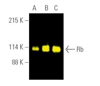
- Rb Antibody (IF8) is recommended for detection of Rb p110 of mouse, rat and human origin by WB, IP, IF, IHC(P) and FCM
- Anti-Rb Antibody (IF8) is available conjugated to agarose for IP; HRP for WB, IHC(P) and ELISA; and to either phycoerythrin or FITC for IF, IHC(P) and FCM
- also available conjugated to Alexa Fluor® 488, Alexa Fluor® 546, Alexa Fluor® 594 or Alexa Fluor® 647 for WB (RGB), IF, IHC(P) and FCM, and for use with RGB fluorescent imaging systems, such as iBright™ FL1000, FluorChem™, Typhoon, Azure and other comparable systems
- also available conjugated to Alexa Fluor® 680 or Alexa Fluor® 790 for WB (NIR), IF and FCM; for use with Near-Infrared (NIR) detection systems, such as LI-COR®Odyssey®, iBright™ FL1000, FluorChem™, Typhoon, Azure and other comparable systems
- also available conjugated to either TRITC or Alexa Fluor® 405 for IF, IHC(P) and FCM
- m-IgG Fc BP-HRP, m-IgG1 BP-HRP and m-IgGκ BP-HRP are the preferred secondary detection reagents for Rb Antibody (IF8) for WB and IHC(P) applications. These reagents are now offered in bundles with Rb Antibody (IF8) (see ordering information below).

QUICK LINKS
SEE ALSO...
Rb Antibody (IF8) is a mouse monoclonal IgG1 kappa light chain antibody that detects the Rb protein (also called retinoblastoma (RB) transcriptional corepressor 1 or RB1) of mouse, rat and human origin by western blotting, immunoprecipitation, immunofluorescence, immunohistochemistry on paraffin embedded sections, and flow cytometry. Rb monoclonal antibody (IF8) is available as both non-conjugated form, as well as multiple conjugated forms, including agarose, horseradish peroxidase, phycoerythrin, fluorescein isothiocyanate, and multiple Alexa Fluor® conjugates. Pediatric cancer retinoblastoma and formation of other human tumors can be attributed to mutations in the retinoblastoma tumor suppressor gene. Rb protein regulates differentiation, apoptosis and cell cycle control by coordinating the cell cycle, at G1/S, with transcriptional machinery that includes the heterodimeric E2F family. During G1, cyclin D (D1, D2, D3)-dependent kinase-mediated phosphorylation of Rb at Ser 795 marks the conversion of Rb from a transcriptionally repressive, hypophosphorylated state to an inactive, phosphorylated state, which may be sustained through mitosis by differential phosphorylation of up to 16 putative serine or threonine residues, including Ser 249/Thr 252, Thr 373, Thr 356, Ser 780, Ser 807/Ser 811 and Thr 821/Thr 826. Hypophosphorylated Rb represses transcription of genes controlling cell cycle through direct protein-protein interactions, by binding and inactivating promoters of transcription factors, and through recruitment of histone deacetylase, which deacetylates promoter regions and enhances nucleosome formation, thereby masking transcription enhancing cis elements. Anti-Rb monoclonal antibody (IF8) is recommended for researchers investigating cell cycle regulation and molecular mechanisms underlying cancer development.
Alexa Fluor® is a trademark of Molecular Probes Inc., OR., USA
LI-COR® and Odyssey® are registered trademarks of LI-COR Biosciences
Rb Antibody (IF8) References:
- Discovery of a regulatory motif that controls the exposure of specific upstream cyclin-dependent kinase sites that determine both conformation and growth suppressing activity of pRb. | Driscoll, B., et al. 1999. J Biol Chem. 274: 9463-71. PMID: 10092628
- Glutamic acid mutagenesis of retinoblastoma protein phosphorylation sites has diverse effects on function. | Barrientes, S., et al. 2000. Oncogene. 19: 562-70. PMID: 10698526
- Transforming growth factor beta inhibits the phosphorylation of pRB at multiple serine/threonine sites and differentially regulates the formation of pRB family-E2F complexes in human myeloid leukemia cells. | Hu, X., et al. 2000. Biochem Biophys Res Commun. 276: 930-9. PMID: 11027571
- The role of the retinoblastoma protein (Rb) in the nuclear localization of BAG-1: implications for colorectal tumour cell survival. | Clemo, NK., et al. 2005. Biochem Soc Trans. 33: 676-8. PMID: 16042572
- Retinoblastoma protein (RB) interacts with E2F3 to control terminal differentiation of Sertoli cells. | Rotgers, E., et al. 2014. Cell Death Dis. 5: e1274. PMID: 24901045
- Phosphorylation of the Retinoblastoma protein (Rb) on serine-807 is required for association with Bax. | Antonucci, LA., et al. 2014. Cell Cycle. 13: 3611-7. PMID: 25483096
- Retinoblastoma protein as an intrinsic BRD4 inhibitor modulates small molecule BET inhibitor sensitivity in cancer. | Ding, D., et al. 2022. Nat Commun. 13: 6311. PMID: 36274096
- The retinoblastoma protein and cell cycle control. | Weinberg, RA. 1995. Cell. 81: 323-30. PMID: 7736585
- Direct transcriptional repression by pRB and its reversal by specific cyclins. | Bremner, R., et al. 1995. Mol Cell Biol. 15: 3256-65. PMID: 7760821
- Cancer cell cycles. | Sherr, CJ. 1996. Science. 274: 1672-7. PMID: 8939849
- Cyclin D1/Cdk4 regulates retinoblastoma protein-mediated cell cycle arrest by site-specific phosphorylation. | Connell-Crowley, L., et al. 1997. Mol Biol Cell. 8: 287-301. PMID: 9190208
- Rb interacts with histone deacetylase to repress transcription. | Luo, RX., et al. 1998. Cell. 92: 463-73. PMID: 9491888
Ordering Information
| Product Name | Catalog # | UNIT | Price | Qty | FAVORITES | |
Rb Antibody (IF8) | sc-102 | 200 µg/ml | $322.00 | |||
Rb Antibody (IF8): m-IgG Fc BP-HRP Bundle | sc-528097 | 200 µg Ab; 10 µg BP | $361.00 | |||
Rb Antibody (IF8): m-IgGκ BP-HRP Bundle | sc-520395 | 200 µg Ab, 40 µg BP | $361.00 | |||
Rb Antibody (IF8): m-IgG1 BP-HRP Bundle | sc-542756 | 200 µg Ab; 20 µg BP | $361.00 | |||
Rb Antibody (IF8) AC | sc-102 AC | 500 µg/ml, 25% agarose | $424.00 | |||
Rb Antibody (IF8) HRP | sc-102 HRP | 200 µg/ml | $322.00 | |||
Rb Antibody (IF8) FITC | sc-102 FITC | 200 µg/ml | $336.00 | |||
Rb Antibody (IF8) PE | sc-102 PE | 200 µg/ml | $349.00 | |||
Rb Antibody (IF8) Alexa Fluor® 488 | sc-102 AF488 | 200 µg/ml | $364.00 | |||
Rb Antibody (IF8) Alexa Fluor® 546 | sc-102 AF546 | 200 µg/ml | $364.00 | |||
Rb Antibody (IF8) Alexa Fluor® 594 | sc-102 AF594 | 200 µg/ml | $364.00 | |||
Rb Antibody (IF8) Alexa Fluor® 647 | sc-102 AF647 | 200 µg/ml | $364.00 | |||
Rb Antibody (IF8) Alexa Fluor® 680 | sc-102 AF680 | 200 µg/ml | $364.00 | |||
Rb Antibody (IF8) Alexa Fluor® 790 | sc-102 AF790 | 200 µg/ml | $364.00 | |||
Rb Antibody (IF8) TRITC | sc-102 TRITC | 200 µg/ml | $326.00 | |||
Rb Antibody (IF8) Alexa Fluor® 405 | sc-102 AF405 | 200 µg/ml | $364.00 |